Movie
Movie Controller
 Structure viewers
 About Yorodumi Papers


+Search query
-Structure paper
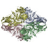
| Title | Structure of β-galactosidase at 3.2-Å resolution obtained by cryo-electron microscopy. |
|---|---|
| Journal, issue, pages | Proc Natl Acad Sci U S A, Vol. 111, Issue 32, Page 11709-11714, Year 2014 |
| Publish date | Aug 12, 2014 |
 Authors Authors | Alberto Bartesaghi / Doreen Matthies / Soojay Banerjee / Alan Merk / Sriram Subramaniam /  |
| PubMed Abstract | We report the solution structure of Escherichia coli β-galactosidase (∼465 kDa), solved at ∼3.2-Å resolution by using single-particle cryo-electron microscopy (cryo-EM). Densities for most side ...We report the solution structure of Escherichia coli β-galactosidase (∼465 kDa), solved at ∼3.2-Å resolution by using single-particle cryo-electron microscopy (cryo-EM). Densities for most side chains, including those of residues in the active site, and a catalytic Mg(2+) ion can be discerned in the map obtained by cryo-EM. The atomic model derived from our cryo-EM analysis closely matches the 1.7-Å crystal structure with a global rmsd of ∼0.66 Å. There are significant local differences throughout the protein, with clear evidence for conformational changes resulting from contact zones in the crystal lattice. Inspection of the map reveals that although densities for residues with positively charged and neutral side chains are well resolved, systematically weaker densities are observed for residues with negatively charged side chains. We show that the weaker densities for negatively charged residues arise from their greater sensitivity to radiation damage from electron irradiation as determined by comparison of density maps obtained by using electron doses ranging from 10 to 30 e(-)/Å(2). In summary, we establish that it is feasible to use cryo-EM to determine near-atomic resolution structures of protein complexes (<500 kDa) with low symmetry, and that the residue-specific radiation damage that occurs with increasing electron dose can be monitored by using dose fractionation tools available with direct electron detector technology. |
 External links External links | Proc Natl Acad Sci U S A / PubMed:25071206 / PubMed Central |
| Methods | EM (single particle) |
| Resolution | 3.2 Å |
| Structure data | |
| Chemicals |  ChemComp-MG: |
| Source |
|
 Keywords Keywords |  HYDROLASE / hydrolase enzyme / homo-tetramer / protein complex / atomic resolution cryo-electron microscopy / direct electron detectors / single-particle cryo-EM / 3D reconstruction HYDROLASE / hydrolase enzyme / homo-tetramer / protein complex / atomic resolution cryo-electron microscopy / direct electron detectors / single-particle cryo-EM / 3D reconstruction |