 Movie
Movie Controller
 Structure viewers
 About Yorodumi Papers


+Search query
-Structure paper
| Title | Three-dimensional architecture of the rod sensory cilium and its disruption in retinal neurodegeneration. |
|---|---|
| Journal, issue, pages | Cell, Vol. 151, Issue 5, Page 1029-1041, Year 2012 |
| Publish date | Nov 21, 2012 |
 Authors Authors | Jared C Gilliam / Juan T Chang / Ivette M Sandoval / Youwen Zhang / Tiansen Li / Steven J Pittler / Wah Chiu / Theodore G Wensel /  |
| PubMed Abstract | Defects in primary cilia lead to devastating disease because of their roles in sensation and developmental signaling but much is unknown about ciliary structure and mechanisms of their formation and ...Defects in primary cilia lead to devastating disease because of their roles in sensation and developmental signaling but much is unknown about ciliary structure and mechanisms of their formation and maintenance. We used cryo-electron tomography to obtain 3D maps of the connecting cilium and adjacent cellular structures of a modified primary cilium, the rod outer segment, from wild-type and genetically defective mice. The results reveal the molecular architecture of the cilium and provide insights into protein functions. They suggest that the ciliary rootlet is involved in cellular transport and stabilizes the axoneme. A defect in the BBSome membrane coat caused defects in vesicle targeting near the base of the cilium. Loss of the proteins encoded by the Cngb1 gene disrupted links between the disk and plasma membranes. The structures of the outer segment membranes support a model for disk morphogenesis in which basal disks are enveloped by the plasma membrane. |
 External links External links | Cell / PubMed:23178122 / PubMed Central |
| Methods | EM (tomography) |
| Structure data | 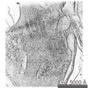 EMDB-5477: 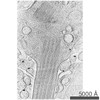 EMDB-5478: 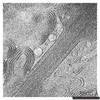 EMDB-5479: 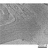 EMDB-5480: 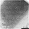 EMDB-5481: 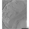 EMDB-5482: 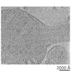 EMDB-5483: 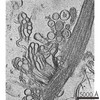 EMDB-5484: |
| Source |
|

 Mus musculus (house mouse)