Movie
Movie Controller
 Structure viewers
 About Yorodumi Papers


+Search query
-Structure paper
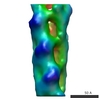
| Title | Molecular structure of the ParM polymer and the mechanism leading to its nucleotide-driven dynamic instability. |
|---|---|
| Journal, issue, pages | EMBO J, Vol. 27, Issue 3, Page 570-579, Year 2008 |
| Publish date | Feb 6, 2008 |
 Authors Authors | David Popp / Akihiro Narita / Toshiro Oda / Tetsuro Fujisawa / Hiroshi Matsuo / Yasushi Nitanai / Mitsusada Iwasa / Kayo Maeda / Hirofumi Onishi / Yuichiro Maéda /  |
| PubMed Abstract | ParM is a prokaryotic actin homologue, which ensures even plasmid segregation before bacterial cell division. In vivo, ParM forms a labile filament bundle that is reminiscent of the more complex ...ParM is a prokaryotic actin homologue, which ensures even plasmid segregation before bacterial cell division. In vivo, ParM forms a labile filament bundle that is reminiscent of the more complex spindle formed by microtubules partitioning chromosomes in eukaryotic cells. However, little is known about the underlying structural mechanism of DNA segregation by ParM filaments and the accompanying dynamic instability. Our biochemical, TIRF microscopy and high-pressure SAX observations indicate that polymerization and disintegration of ParM filaments is driven by GTP rather than ATP and that ParM acts as a GTP-driven molecular switch similar to a G protein. Image analysis of electron micrographs reveals that the ParM filament is a left-handed helix, opposed to the right-handed actin polymer. Nevertheless, the intersubunit contacts are similar to those of actin. Our atomic model of the ParM-GMPPNP filament, which also fits well to X-ray fibre diffraction patterns from oriented gels, can explain why after nucleotide release, large conformational changes of the protomer lead to a breakage of intra- and interstrand interactions, and thus to the observed disintegration of the ParM filament after DNA segregation. |
 External links External links | EMBO J / PubMed:18188150 / PubMed Central |
| Methods | EM (helical sym.) / X-ray diffraction |
| Resolution | 1.9 - 23.0 Å |
| Structure data | EMDB-1470: Molecular structure of the ParM polymer and the mechanism leading to its nucleotide-driven dynamic instability  PDB-2zgy:  PDB-2zgz: |
| Chemicals |  ChemComp-MG:  ChemComp-GDP:  ChemComp-HOH:  ChemComp-GNP:  ChemComp-ADP: |
| Source |
|
 Keywords Keywords |  STRUCTURAL PROTEIN / PARM / Plasmid / Plasmid partition / CELL CYCLE/PROTEIN FIBRIL / CELL CYCLE-PROTEIN FIBRIL COMPLEX STRUCTURAL PROTEIN / PARM / Plasmid / Plasmid partition / CELL CYCLE/PROTEIN FIBRIL / CELL CYCLE-PROTEIN FIBRIL COMPLEX |