Christiane Schaffitzel / Miro Oswald / Imre Berger / Takashi Ishikawa / Jan Pieter Abrahams / Henk K Koerten / Roman I Koning / Nenad Ban /
PubMed Abstract
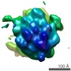
The prokaryotic signal recognition particle (SRP) targets membrane proteins into the inner membrane. It binds translating ribosomes and screens the emerging nascent chain for a hydrophobic signal ...The prokaryotic signal recognition particle (SRP) targets membrane proteins into the inner membrane. It binds translating ribosomes and screens the emerging nascent chain for a hydrophobic signal sequence, such as the transmembrane helix of inner membrane proteins. If such a sequence emerges, the SRP binds tightly, allowing the SRP receptor to lock on. This assembly delivers the ribosome-nascent chain complex to the protein translocation machinery in the membrane. Using cryo-electron microscopy and single-particle reconstruction, we obtained a 16 A structure of the Escherichia coli SRP in complex with a translating E. coli ribosome containing a nascent chain with a transmembrane helix anchor. We also obtained structural information on the SRP bound to an empty E. coli ribosome. The latter might share characteristics with a scanning SRP complex, whereas the former represents the next step: the targeting complex ready for receptor binding. High-resolution structures of the bacterial ribosome and of the bacterial SRP components are available, and their fitting explains our electron microscopic density. The structures reveal the regions that are involved in complex formation, provide insight into the conformation of the SRP on the ribosome and indicate the conformational changes that accompany high-affinity SRP binding to ribosome nascent chain complexes upon recognition of the signal sequence.
EMDB-1250: Structure of the E. coli signal recognition particle bound to a translating ribosome. PDB-2iy3: Structure of the E. Coli Signal Regognition Particle Method: EM (single particle) / Resolution: 15.5 Å
EMDB-1251: Structure of the E. coli signal recognition particle bound to a translating ribosome. PDB-2iy3: Structure of the E. Coli Signal Regognition Particle Method: EM (single particle) / Resolution: 20.0 Å
Source
escherichia coli (E. coli)
thermus aquaticus (bacteria)
sulfolobus solfataricus (archaea)
synthetic construct (others)
Keywords
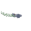
RNA-BINDING / RNA-BINDING PROTEIN COMPLEX / SIGNAL RECOGNITION PARTICLE
+
About Yorodumi Papers
-
News
-
Feb 9, 2022. New format data for meta-information of EMDB entries
New format data for meta-information of EMDB entries
Version 3 of the EMDB header file is now the official format.
The previous official version 1.9 will be removed from the archive.
In the structure databanks used in Yorodumi, some data are registered as the other names, "COVID-19 virus" and "2019-nCoV". Here are the details of the virus and the list of structure data.
Jan 31, 2019. EMDB accession codes are about to change! (news from PDBe EMDB page)
EMDB accession codes are about to change! (news from PDBe EMDB page)
The allocation of 4 digits for EMDB accession codes will soon come to an end. Whilst these codes will remain in use, new EMDB accession codes will include an additional digit and will expand incrementally as the available range of codes is exhausted. The current 4-digit format prefixed with “EMD-” (i.e. EMD-XXXX) will advance to a 5-digit format (i.e. EMD-XXXXX), and so on. It is currently estimated that the 4-digit codes will be depleted around Spring 2019, at which point the 5-digit format will come into force.
The EM Navigator/Yorodumi systems omit the EMD- prefix.
Related info.:Q: What is EMD? / ID/Accession-code notation in Yorodumi/EM Navigator
 Movie
Movie Controller
 Structure viewers
 About Yorodumi Papers


 Authors
 External links
 Keywords
 SIGNAL RECOGNITION PARTICLE