 ムービー
ムービー コントローラー
 構造ビューア
 EMN検索について


-検索条件
-検索結果
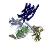
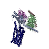
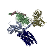
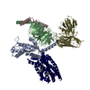
検索 (著者・登録者: x. & yu)の結果1,603件中、1から50件目までを表示しています
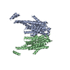
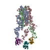
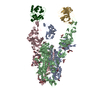
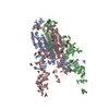
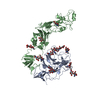
PDB-8xit: 
Cryo-EM structure of sheep VMAT2 dimer in an atypical fold
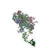
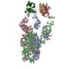
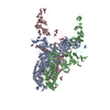
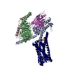
PDB-8xiu:
Cryo-EM structure of a frog VMAT2 in an apo conformation

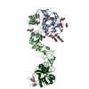
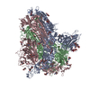
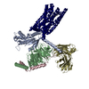
PDB-8y6v:
Near-atomic structure of icosahedrally averaged jumbo bacteriophage PhiKZ capsid
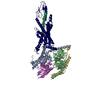
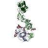
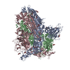
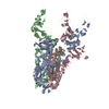
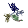
PDB-8xva:
Human TOM complex with whole Tom20
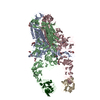
PDB-8yw5:
Cryo-EM structure of the retatrutide-bound human GCGR-Gs complex

PDB-8w2f:
Plasmodium falciparum 20S proteasome bound to an inhibitor

PDB-8vt2:
cryo-EM structure of HMPV (MPV-2c)
PDB-8vt3:
cryo-EM structure of HMPV (MPV-2cREKR)

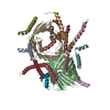
PDB-8y36:
cryo-EM structure of Staphylococcus aureus(ATCC 29213) 50S ribosome in complex with MCX-190.

PDB-8y37:
Cryo-EM structure of Staphylococcus aureus (15B196) 50S ribosome in complex with MCX-190.

PDB-8y38:
Cryo-EM structure of Staphylococcus aureus 70S ribosome (strain 15B196) in complex with MCX-190.

PDB-8y39:
cryo-EM structure of Staphylococcus aureus(ATCC 29213) 70S ribosome in complex with MCX-190.
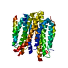
PDB-8wlu:
Cryo-EM structure of bat RsSHC014 spike glycoprotein
PDB-8wly:
Cryo-EM structure of bat WIV1 spike glycoprotein
PDB-8wlz:
Cryo-EM structure of the WIV1 S-hACE2 complex

PDB-8wq0:
Cryo-EM structure of WIV1 spike glycoprotein (the closed state)
PDB-8y7x:
Structure of HCoV-HKU1A spike in the functionally anchored-3up conformation with 3TMPRSS2
PDB-8y7y:
Local structure of HCoV-HKU1A spike in complex with TMPRSS2 and glycan
PDB-8y87:
Structure of HCoV-HKU1C spike in the functionally anchored-1up conformation with 1TMPRSS2
PDB-8y88:
Structure of HCoV-HKU1C spike in the functionally anchored-2up conformation with 2TMPRSS2
PDB-8y89:
Structure of HCoV-HKU1C spike in the functionally anchored-3up conformation with 2TMPRSS2
PDB-8y8a:
Structure of HCoV-HKU1C spike in the functionally anchored-3up conformation with 3TMPRSS2
PDB-8y8b:
Local structure of HCoV-HKU1C spike in complex with TMPRSS2 and glycan
PDB-8y8c:
Structure of HCoV-HKU1C spike in the inactive-closed conformation

PDB-8y8d:
Structure of HCoV-HKU1C spike in the inactive-1up conformation
PDB-8y8e:
Structure of HCoV-HKU1C spike in the inactive-2up conformation
PDB-8y8f:
Structure of HCoV-HKU1C spike in the glycan-activated-closed conformation

PDB-8y8g:
Structure of HCoV-HKU1C spike in the glycan-activated-1up conformation
PDB-8y8h:
Structure of HCoV-HKU1C spike in the glycan-activated-2up conformation
PDB-8y8i:
Structure of HCoV-HKU1C spike in the glycan-activated-3up conformation
PDB-8y8j:
Local structure of HCoV-HKU1C spike in complex with glycan

PDB-8xpq:
Structure of the sea urchin spSLC9C1 in state-2 w/o cAMP dimer

PDB-8xq4:
Structure of the sea urchin spSLC9C1 in state-2 w/o cAMP protomer

PDB-8xq7:
Structure of the sea urchin spSLC9C1 in state-1 w/ cAMP dimer

PDB-8xq8:
Structure of the sea urchin spSLC9C1 in state-1 w/ cAMP protomer

PDB-8xq9:
Structure of the sea urchin spSLC9C1 in state-2 w/ cAMP dimer

PDB-8xqa:
Structure of the sea urchin spSLC9C1 in state-3 w/ cAMP dimer

PDB-8xki:
A neutralizing nanobody VHH60 against wt SARS-CoV-2

PDB-8zbe:
cryo-EM structure of the octreotide-bound SSTR5-Gi complex

PDB-8zcj:
Cryo-EM structure of the pasireotide-bound SSTR5-Gi complex
PDB-8xql:
Structure of human class T GPCR TAS2R14-miniGs/gust complex with Aristolochic acid A.
PDB-8xqn:
Structure of human class T GPCR TAS2R14-DNGi complex with Aristolochic acid A.
PDB-8xqo:
Structure of human class T GPCR TAS2R14-Gi complex with Aristolochic acid A.
PDB-8xqp:
Structure of human class T GPCR TAS2R14-Gustducin complex with Aristolochic acid A.
PDB-8xqr:
Structure 2 of human class T GPCR TAS2R14-miniGs/gust complex with Flufenamic acid.
PDB-8xqs:
Structure of human class T GPCR TAS2R14-DNGi complex with Flufenamic acid.
PDB-8xqt:
Structure of human class T GPCR TAS2R14-Gi complex.
PDB-8yky:
Structure of human class T GPCR TAS2R14-Ggustducin complex with agonist 28.1

PDB-8k10:
SID1 transmembrane family member 2

PDB-8k11:
SID1 transmembrane family member 2
ページ:
 wwPDBはEMDBデータモデルのバージョン3へ移行します
