 Movie
Movie Controller
 Structure viewers
 About EMN search


-Search query
-Search result
Showing all 22 items for (author: schorb & m)
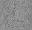
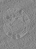
EMDB-16128: 
Tomogram of a late endosome of A549 cell infected with influenza A virus.
Method: electron tomography / : Klein S, Chlanda P
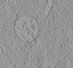
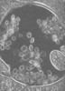
EMDB-16129:
Tomogram of a late endosome of A549 cell infected with influenza A virus (Figure 6C).
Method: electron tomography / : Klein S, Chlanda P

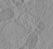
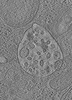
EMDB-16130:
Tomogram of a late endosome of A549 cell infected with influenza A virus (Figure 6E)
Method: electron tomography / : Klein S, Chlanda P
EMDB-16131:
Tomogram of a late endosome of A549 cell infected with influenza A virus (Figure 6G)
Method: electron tomography / : Klein S, Chlanda P
EMDB-16132:
Tomogram of a late endosome of A549 cell infected with influenza A virus (Figure 6I,K,M)
Method: electron tomography / : Klein S, Chlanda P
EMDB-16133:
Tomogram of a late endosome of A549 cell infected with influenza A virus (Figure 6O)
Method: electron tomography / : Klein S, Chlanda P
EMDB-15705:
Tomogram of a late endosome of A549 cell (Figure 1R)
Method: electron tomography / : Klein S, Chlanda P
EMDB-15707:
Tomogram of a late endosome of A549 cell treated with IFN-beta (Figure 1T)
Method: electron tomography / : Klein S, Chlanda P
EMDB-15708:
Tomogram of a late endosome of A549-IFITM3 cell (Figure 1V)
Method: electron tomography / : Klein S, Chlanda P
EMDB-15131:
Tomogram of a late endosome of A549-IFITM3 cells infected with influenza A virus (Figure S7)
Method: electron tomography / : Klein S, Chlanda P
EMDB-15130:
Tomogram of a late endosome of A549-IFITM3 cells infected with influenza A virus (Figure 4E)
Method: electron tomography / : Klein S, Chlanda P
EMDB-15132:
Tomogram of a late endosome of A549-IFITM3 cells infected with influenza A virus (Figure S8)
Method: electron tomography / : Klein S, Chlanda P

EMDB-15133:
Tomogram of a late endosome of A549-IFITM3 cells infected with influenza A virus (Figure S9)
Method: electron tomography / : Klein S, Chlanda P

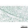
EMDB-12940:
SARS-CoV-2-Induced Reshaping of Subcellular Morphologies
Method: electron tomography / : Cortese M, Schwab Y, Bartenschlager R, Schorb M
EMDB-3820:
Electron tomographic slices of the nuclear envelope of HeLa cell in interphase
Method: electron tomography / : Otsuka S, Ellenberg J

PDB-4d1k:
Cryo-electron microscopy of tubular arrays of HIV-1 Gag resolves structures essential for immature virus assembly.
Method: helical / : Bharat TAM, Castillo-Menendez LR, Hagen WJH, Lux V, Igonet S, Schorb M, Schur FKM, Krauesslich HG, Briggs JAG
EMDB-2638:
Cryo-electron microscopy of tubular arrays of HIV-1 Gag resolves structures essential for immature virus assembly.
Method: helical / : Bharat TAM, Castillo-Menendez LR, Hagen WH, Lux V, Igonet S, Schorb M, Schur FKM, Krauesslich HG, Briggs JAG
EMDB-2084:
Structures from COPI-coated vesicles: triad
Method: subtomogram averaging / : Faini M, Prinz S, Beck R, Schorb M, Riches JD, Bacia K, Brugger B, Wieland FT, Briggs JAG
EMDB-2085:
Structures from COPI-coated vesicles: three-corner connection
Method: subtomogram averaging / : Faini M, Prinz S, Beck R, Schorb M, Riches JD, Bacia K, Brugger B, Wieland FT, Briggs JAG
EMDB-2086:
Structures from COPI-coated vesicles: three-edge connection
Method: subtomogram averaging / : Faini M, Prinz S, Beck R, Schorb M, Riches JD, Bacia K, Brugger B, Wieland FT, Briggs JAG
EMDB-2087:
Structures from COPI-coated vesicles: single two-corner connection
Method: subtomogram averaging / : Faini M, Prinz S, Beck R, Schorb M, Riches JD, Bacia K, Brugger B, Wieland FT, Briggs JAG
EMDB-2088:
Structures from COPI-coated vesicles: paired two-corner connection
Method: subtomogram averaging / : Faini M, Prinz S, Beck R, Schorb M, Riches JD, Bacia K, Brugger B, Wieland FT, Briggs JAG
 wwPDB to switch to version 3 of the EMDB data model
