 Movie
Movie Controller
 Structure viewers
 About EMN search


-Search query
-Search result
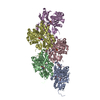
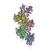
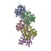
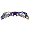
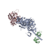
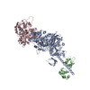
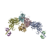
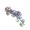
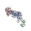
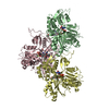
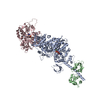
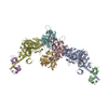
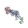
Showing 1 - 50 of 130 items for (author: raunser, & s.)

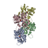
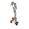
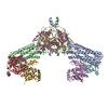
PDB-8rty: 
Structure of the F-actin barbed end bound by Cdc12 and profilin (ring complex) at a resolution of 6.3 Angstrom
Method: single particle / : Oosterheert W, Boiero Sanders M, Funk J, Prumbaum D, Raunser S, Bieling P
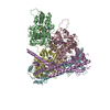
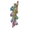
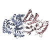
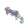
PDB-8ru0:
Structure of the undecorated barbed end of F-actin.
Method: single particle / : Oosterheert W, Boiero Sanders M, Funk J, Prumbaum D, Raunser S, Bieling P
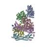
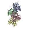
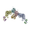
PDB-8ru2:
Structure of the F-actin barbed end bound by formin mDia1
Method: single particle / : Oosterheert W, Boiero Sanders M, Funk J, Prumbaum D, Raunser S, Bieling P
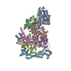
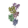
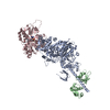
PDB-8rv2:
Structure of the formin INF2 bound to the barbed end of F-actin.
Method: single particle / : Oosterheert W, Boiero Sanders M, Funk J, Prumbaum D, Raunser S, Bieling P

PDB-8s3e:
Structure of rabbit Slo1 in complex with gamma1/LRRC26
Method: single particle / : Redhardt M, Raunser S, Raisch T
PDB-8rtt:
Structure of the formin Cdc12 bound to the barbed end of phalloidin-stabilized F-actin.
Method: single particle / : Oosterheert W, Boiero Sanders M, Funk J, Prumbaum D, Raunser S, Bieling P

PDB-8cpz:
Photorhabdus luminescens TcdA1 prepore-to-pore intermediate, K1179W mutant
Method: single particle / : Nganga PN, Roderer D, Belyy A, Prumbaum D, Raunser S

PDB-8cq0:
Photorhabdus luminescens TcdA1 prepore-to-pore intermediate, K567W K2008W mutant
Method: single particle / : Nganga PN, Roderer D, Belyy A, Prumbaum D, Raunser S

PDB-8cq2:
Photorhabdus luminescens TcdA1 prepore-to-pore intermediate, C16S, C20S, C870S, T1279C mutant
Method: single particle / : Nganga PN, Roderer D, Belyy A, Prumbaum D, Raunser S
PDB-8q5h:
Human KMN network (outer kinetochore)
Method: single particle / : Raisch T, Polley S, Vetter I, Musacchio A, Raunser S

PDB-8p50:
Photorhabdus luminescens Makes caterpillars floppy (Mcf) toxin with the C-terminal deletion in complex with Arf3
Method: single particle / : Belyy A, Heilen P, Hofnagel O, Raunser S

PDB-8p51:
Photorhabdus luminescens Makes caterpillars floppy (Mcf) toxin with the C-terminal deletion
Method: single particle / : Belyy A, Heilen P, Hofnagel O, Raunser S

PDB-8p52:
Photorhabdus luminescens Makes caterpillars floppy (Mcf) toxin
Method: single particle / : Belyy A, Heilen P, Hofnagel O, Raunser S
PDB-8q4g:
Thin filament from FIB milled relaxed left ventricular mouse myofibrils
Method: subtomogram averaging / : Tamborrini D, Wang Z, Wagner T, Tacke S, Stabrin M, Grange M, Kho AL, Bennet P, Rees M, Gautel M, Raunser S

PDB-8q6t:
Helical reconstruction of the relaxed thick filament from FIB milled left ventricular mouse myofibrils
Method: subtomogram averaging / : Tamborrini D, Raunser S

PDB-8oi8:
Cryo-EM structure of ADP-bound, filamentous beta-actin harboring the R183W mutation
Method: single particle / : Oosterheert W, Blanc FEC, Roy A, Belyy A, Hofnagel O, Hummer G, Bieling P, Raunser S
PDB-8oi6:
Cryo-EM structure of the undecorated barbed end of filamentous beta/gamma actin
Method: single particle / : Oosterheert W, Blanc FEC, Roy A, Belyy A, Hofnagel O, Hummer G, Bieling P, Raunser S
PDB-8oid:
Cryo-EM structure of ADP-bound, filamentous beta-actin harboring the N111S mutation
Method: single particle / : Oosterheert W, Blanc FEC, Roy A, Belyy A, Hofnagel O, Hummer G, Bieling P, Raunser S

PDB-8atd:
Wild type hexamer oxalyl-CoA synthetase (OCS)
Method: single particle / : Lill P, Burgi J, Raunser S, Wilmanns M, Gatsogiannis C

PDB-8a2r:
Cryo-EM structure of F-actin in the Mg2+-ADP-BeF3- nucleotide state.
Method: single particle / : Oosterheert W, Klink BU, Belyy A, Pospich S, Raunser S
PDB-8a2s:
Cryo-EM structure of F-actin in the Mg2+-ADP-Pi nucleotide state.
Method: single particle / : Oosterheert W, Klink BU, Belyy A, Pospich S, Raunser S
PDB-8a2t:
Cryo-EM structure of F-actin in the Mg2+-ADP nucleotide state.
Method: single particle / : Oosterheert W, Klink BU, Belyy A, Pospich S, Raunser S

PDB-8a2u:
Cryo-EM structure of F-actin in the Ca2+-ADP-BeF3- nucleotide state.
Method: single particle / : Oosterheert W, Klink BU, Belyy A, Pospich S, Raunser S

PDB-8a2y:
Cryo-EM structure of F-actin in the Ca2+-ADP-Pi nucleotide state.
Method: single particle / : Oosterheert W, Klink BU, Belyy A, Pospich S, Raunser S
PDB-8a2z:
Cryo-EM structure of F-actin in the Ca2+-ADP nucleotide state.
Method: single particle / : Oosterheert W, Klink BU, Belyy A, Pospich S, Raunser S

PDB-7z7h:
Structure of P. luminescens TccC3-F-actin complex
Method: single particle / : Belyy A, Raunser S


PDB-7zgu:
Human NLRP3-deltaPYD hexamer
Method: single particle / : Raisch T, Machtens DA, Bresch IB, Eberhage J, Prumbaum D, Reubold TF, Raunser S, Eschenburg S
PDB-7qpg:
Human RZZ kinetochore corona complex.
Method: single particle / : Raisch T, Ciossani G, d'Amico E, Cmetowski V, Carmignani S, Maffini S, Merino F, Wohlgemuth S, Vetter IR, Raunser S, Musacchio A

PDB-7qim:
In situ structure of nebulin bound to actin filament in skeletal sarcomere
Method: subtomogram averaging / : Wang Z, Grange M, Pospich S, Wagner T, Kho AL, Gautel M, Raunser S
PDB-7nd2:
Cryo-EM structure of the human FERRY complex
Method: single particle / : Quentin D, Klink BU, Raunser S

PDB-7qin:
In situ structure of actomyosin complex in skeletal sarcomere
Method: subtomogram averaging / : Wang Z, Grange M, Pospich S, Wagner T, Kho AL, Gautel M, Raunser S

PDB-7qio:
Homology model of myosin neck domain in skeletal sarcomere
Method: subtomogram averaging / : Wang Z, Grange M, Pospich S, Wagner T, Kho AL, Gautel M, Raunser S
PDB-7qla:
Structure of the Rab GEF complex Mon1-Ccz1
Method: single particle / : Klink BU, Herrmann E, Antoni C, Langemeyer L, Kiontke S, Gatsogiannis C, Ungermann C, Raunser S, Kuemmel D

PDB-7q97:
Structure of the bacterial type VI secretion system effector RhsA.
Method: single particle / : Guenther P, Quentin D, Ahmad S, Sachar K, Gatsogiannis C, Whitney JC, Raunser S

PDB-7plt:
Cryo-EM structure of the actomyosin-V complex in the rigor state (central 1er)
Method: helical / : Pospich S, Sweeney HL, Houdusse A, Raunser S
PDB-7plu:
Cryo-EM structure of the actomyosin-V complex in the rigor state (central 3er/2er)
Method: helical / : Pospich S, Sweeney HL, Houdusse A, Raunser S
PDB-7plv:
Cryo-EM structure of the actomyosin-V complex in the rigor state (central 1er, class 1)
Method: helical / : Pospich S, Sweeney HL, Houdusse A, Raunser S
PDB-7plw:
Cryo-EM structure of the actomyosin-V complex in the rigor state (central 1er, class 2)
Method: helical / : Pospich S, Sweeney HL, Houdusse A, Raunser S

PDB-7plx:
Cryo-EM structure of the actomyosin-V complex in the rigor state (central 1er, class 4)
Method: helical / : Pospich S, Sweeney HL, Houdusse A, Raunser S
PDB-7ply:
Cryo-EM structure of the actomyosin-V complex in the rigor state (central 1er, young JASP-stabilized F-actin)
Method: helical / : Pospich S, Sweeney HL, Houdusse A, Raunser S
PDB-7plz:
Cryo-EM structure of the actomyosin-V complex in the rigor state (central 3er/2er, young JASP-stabilized F-actin)
Method: helical / : Pospich S, Sweeney HL, Houdusse A, Raunser S

PDB-7pm0:
Cryo-EM structure of the actomyosin-V complex in the rigor state (central 1er, young JASP-stabilized F-actin, class 1)
Method: helical / : Pospich S, Sweeney HL, Houdusse A, Raunser S
PDB-7pm1:
Cryo-EM structure of the actomyosin-V complex in the rigor state (central 1er, young JASP-stabilized F-actin, class 2)
Method: helical / : Pospich S, Sweeney HL, Houdusse A, Raunser S
PDB-7pm2:
Cryo-EM structure of the actomyosin-V complex in the rigor state (central 1er, young JASP-stabilized F-actin, class 4)
Method: helical / : Pospich S, Sweeney HL, Houdusse A, Raunser S
PDB-7pm3:
Cryo-EM structure of young JASP-stabilized F-actin (central 3er)
Method: helical / : Pospich S, Sweeney HL, Houdusse A, Raunser S
PDB-7pm5:
Cryo-EM structure of the actomyosin-V complex in the strong-ADP state (central 1er)
Method: helical / : Pospich S, Sweeney HL, Houdusse A, Raunser S
PDB-7pm6:
Cryo-EM structure of the actomyosin-V complex in the strong-ADP state (central 3er/2er)
Method: helical / : Pospich S, Sweeney HL, Houdusse A, Raunser S
PDB-7pm7:
Cryo-EM structure of the actomyosin-V complex in the strong-ADP state (central 1er, class 2)
Method: helical / : Pospich S, Sweeney HL, Houdusse A, Raunser S
PDB-7pm8:
Cryo-EM structure of the actomyosin-V complex in the strong-ADP state (central 1er, class 3)
Method: helical / : Pospich S, Sweeney HL, Houdusse A, Raunser S
Pages:
 wwPDB to switch to version 3 of the EMDB data model
