 Movie
Movie Controller
 Structure viewers
 About EMN search


-Search query
-Search result
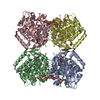
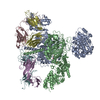
Showing 1 - 50 of 3,398 items for (author: l. & li)

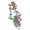
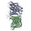
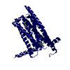
PDB-8y6v: 
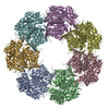
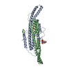
Near-atomic structure of icosahedrally averaged jumbo bacteriophage PhiKZ capsid
Method: single particle / : Yang Y, Shao Q, Guo M, Han L, Zhao X, Wang A, Li X, Wang B, Pan J, Chen Z, Fokine A, Sun L, Fang Q
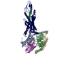
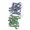
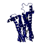
PDB-8u39:
Structure of Human Mitochondrial Chaperonin V72I mutant
Method: single particle / : Chen L, Wang J
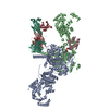
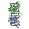
PDB-8kdm:
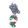
Structure of SARS-CoV Spike protein complexed with antibody PW5-5
Method: single particle / : Sun L, Mao Q, Wang Y

PDB-8kdr:
The local refined map of SARS-CoV-2 XBB Variant Spike protein complexed with antibody PW5-535
Method: single particle / : Sun L, Mao Q, Wang Y
PDB-8kds:
Trimer state of SARS-CoV Spike protein complexed with antibody PW5-535
Method: single particle / : Sun L, Mao Q, Wang Y

PDB-8kdt:
The local refined map of SARS-CoV Spike protein complexed with antibody PW5-5
Method: single particle / : Sun L, Mao Q, Wang Y
PDB-8kej:
Monomer state of SARS-CoV-2 XBB Variant Spike protein trimer complexed with antibody PW5-5
Method: single particle / : Sun L, Mao Q, Wang Y

PDB-8kek:
Monomer state of SARS-CoV Spike protein complexed with antibody PW5-535
Method: single particle / : Sun L, Mao Q, Wang Y
PDB-8keo:
Structure of SARS-CoV-2 Omicron BA.1 Spike complexed with antibody PW5-570
Method: single particle / : Sun L, Mao Q, Wang Y

PDB-8kep:
The local refined map of SARS-CoV-2 Omicron BA.1 Spike complexed with antibody PW5-570
Method: single particle / : Sun L, Mao Q, Wang Y

PDB-8keq:
State 1 of SARS-CoV-2 XBB Variant Spike protein trimer complexed with antibody PW5-5
Method: single particle / : Sun L, Mao Q, Wang Y

PDB-8ker:
Structure of SARS-CoV-2 XBB Variant Spike protein complexed with broadly neutralizing antibody PW5-535
Method: single particle / : Sun L, Mao Q, Wang Y

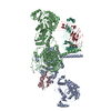
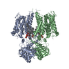
PDB-9f2k:
Myo-inositol-1-phosphate synthase from Thermochaetoides thermophila in complex with NAD
Method: single particle / : Traeger TK, Kyrilis FL, Hamdi F, Kastritis PL
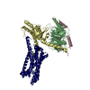
PDB-8yw5:
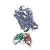
Cryo-EM structure of the retatrutide-bound human GCGR-Gs complex
Method: single particle / : Li WZ, Zhou QT, Cong ZT, Yuan QN, Li WX, Zhao FH, Xu HE, Zhao LH, Yang DH, Wang MW


PDB-8x7i:
Cryo-EM structures of RNF168/UbcH5c-Ub in complex with H2AK13Ub nucleosomes determined by intein-based E2-Ub-NCP conjugation strategy
Method: single particle / : Ai HS, Tong ZB, Deng ZH, Pan M, Liu L

PDB-8x7j:
Cryo-EM structures of RNF168/UbcH5c-Ub/nucleosomes complex determined by activity-based chemical trapping strategy
Method: single particle / : Ai HS, Tong ZB, Deng ZH, Pan M, Liu L

PDB-8x7k:
Cryo-EM structures of RNF168/UbcH5c-Ub in complex with H2AK13Ub nucleosomes determined by activity-based chemical trapping strategy (adjacent H2AK13/15 dual-monoubiquitination)
Method: single particle / : Ai HS, Tong ZB, Deng ZH, Pan M, Liu L

PDB-8tpw:
Crosslinked 6-deoxyerythronolide B synthase (DEBS) Module 3 in complex with antibody fragment 1B2: cis-oriented 1B2 and ACP
Method: single particle / : Cogan DP, Soohoo AM, Chen M, Brodsky KL, Liu Y, Khosla C
PDB-8tpx:
Crosslinked 6-deoxyerythronolide B synthase (DEBS) Module 3 in complex with antibody fragment 1B2: trans-oriented 1B2 and ACP
Method: single particle / : Cogan DP, Soohoo AM, Chen M, Brodsky KL, Liu Y, Khosla C
PDB-8y85:
Human AE3 with NaHCO3- and DIDS
Method: single particle / : Jian L, Zhang Q, Yao D, Wang Q, Xia Y, Qin A, Cao Y
PDB-8y8k:
The structure of hAE3
Method: single particle / : Jian L, Zhang Q, Yao D, Wang Q, Xia Y, Qin A, Cao Y
PDB-8zle:
hAE3NTD2TMD with PT5,CLR, and Y01
Method: single particle / : Jian L, Zhang Q, Yao D, Wang Q, Xia Y, Qin A, Cao Y
PDB-8yww:
The structure of HKU1-B S protein with bsAb1
Method: single particle / : Xia LY, Zhang YY, Zhou Q
PDB-8ywx:
the complex structure of the H4B6 Fab with the RBD of Omicron BA.5 S protein
Method: single particle / : Xia LY, Zhang YY, Zhou Q
PDB-8xlp:
Structure of inactive Photosystem II associated with CAC antenna from Rhodomonas Salina
Method: single particle / : Si L, Li M


PDB-8zfk:
Caenorhabditis elegans ACR-23 in betaine and monepantel bound state
Method: single particle / : Chen QF, Liu FL, Li TY, Gong HH, Guo F, Liu S
PDB-8zfl:
Caenorhabditis elegans ACR-23 in apo state
Method: single particle / : Chen QF, Liu FL, Li TY, Gong HH, Guo F, Liu S
PDB-8zfm:
Caenorhabditis elegans ACR-23 in betaine bound state
Method: single particle / : Chen QF, Liu FL, Li TY, Gong HH, Guo F, Liu S
PDB-8tko:
KS-AT core of 6-deoxyerythronolide B synthase (DEBS) Module 3 crosslinked with its translocation ACP partner of Module 2
Method: single particle / : Cogan DP, Soohoo AM, Chen M, Brodsky KL, Liu Y, Khosla C
PDB-8qb7:
Pil1 in native eisosome lattice bound to plasma membrane microdomain
Method: single particle / : Kefauver JM, Zou L, Loewith RJ, Defosses A
PDB-8qb8:
Lsp1 in native eisosome lattice bound to plasma membrane microdomain
Method: single particle / : Kefauver JM, Zou L, Loewith RJ, Defosses A
PDB-8qb9:
Helical reconstruction of yeast eisosome protein Pil1 bound to membrane composed of lipid mixture -PIP2/+sterol (DOPC, DOPE, DOPS, cholesterol 30:20:20:30)
Method: helical / : Kefauver JM, Zou L, Desfosses A, Loewith RJ
PDB-8qbb:
Helical reconstruction of yeast eisosome protein Pil1 bound to membrane composed of lipid mixture +PIP2/-sterol (DOPC, DOPE, DOPS, PI(4,5)P2 50:20:20:10)
Method: helical / : Kefauver JM, Zou L, Desfosses A, Loewith RJ
PDB-8qbd:
Helical reconstruction of yeast eisosome protein Pil1 bound to membrane composed of lipid mixture +PIP2/+sterol (DOPC, DOPE, DOPS, cholesterol, PI(4,5)P2 35:20:20:15:10)
Method: helical / : Kefauver JM, Zou L, Desfosses A, Loewith RJ
PDB-8qbe:
Compact state - Pil1 in native eisosome lattice bound to plasma membrane microdomain
Method: single particle / : Kefauver JM, Zou L, Desfosses A, Loewith RJ
PDB-8qbf:
Compact state - Pil1 dimer with lipid headgroups fitted in native eisosome lattice bound to plasma membrane microdomain
Method: single particle / : Kefauver JM, Zou L, Desfosses A, Loewith RJ
PDB-8qbg:
Stretched state - Pil1 in native eisosome lattice bound to plasma membrane microdomain
Method: single particle / : Kefauver JM, Zou L, Desfosses A, Loewith RJ
PDB-9euo:
Outward-open structure of Drosophila dopamine transporter bound to an atypical non-competitive inhibitor
Method: single particle / : Pedersen CN, Yang F, Ita S, Xu Y, Akunuri R, Trampari S, Neumann CMT, Desdorf LM, Schioett B, Salvino JM, Mortensen OV, Nissen P, Shahsavar A
PDB-9eup:
Inhibitor-free outward-open structure of Drosophila dopamine transporter
Method: single particle / : Pedersen CN, Yang F, Ita S, Xu Y, Akunuri R, Trampari S, Neumann CMT, Desdorf LM, Schioett B, Salvino JM, Mortensen OV, Nissen P, Shahsavar A
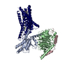
PDB-9iiw:
A local Cryo-EM structure of Bitter taste receptor TAS2R14
Method: single particle / : Yuan Q, Duan J, Tao L, Xu EH
PDB-9iix:
A Cryo-EM structure of Bitter taste receptor TAS2R14 with Ggust
Method: single particle / : Yuan Q, Duan J, Tao L, Xu EH
PDB-9ij9:
A Cryo-EM structure of Bitter taste receptor TAS2R14 with Gi complex
Method: single particle / : Yuan Q, Duan J, Tao L, Xu EH
PDB-9ija:
A local Cryo-EM structure of Bitter taste receptor TAS2R14 with Gi complex
Method: single particle / : Yuan Q, Duan J, Tao L, Xu EH
PDB-8viw:
Cryo-EM structure of heparosan synthase 2 from Pasteurella multocida with polysaccharide in the GlcNAc-T active site
Method: single particle / : Krahn JM, Pedersen LC, Liu J, Stancanelli E, Borgnia M, Vivarette E

PDB-8tjn:
Crosslinked 6-deoxyerythronolide B synthase (DEBS) Module 1 in complex with antibody fragment 1B2: Crosslinked State 1
Method: single particle / : Cogan DP, Soohoo AM, Chen M, Brodsky KL, Liu Y, Khosla C
Pages:
 wwPDB to switch to version 3 of the EMDB data model
