 ムービー
ムービー コントローラー
 構造ビューア
 EMN検索について


-検索条件
-検索結果
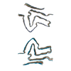
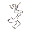
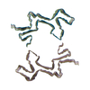
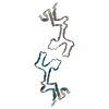
検索 (著者・登録者: g. & schroeder)の結果全49件を表示しています
PDB-8oqi: 
Cryo-EM structure of the wild-type alpha-synuclein fibril.
PDB-8ovk:
Lipidic amyloid-beta(1-40) fibril - polymorph L1
PDB-8ovm:
Lipidic amyloid-beta(1-40) fibril - polymorph L2
PDB-8owd:
Lipidic amyloid-beta(1-40) fibril - polymorph L3
PDB-8owe:
Lipidic amyloid-beta(1-40) fibril - polymorph L2-L3
PDB-8owj:
Lipidic amyloid-beta(1-40) fibril - polymorph L2-L2
PDB-8owk:
Lipidic amyloid-beta(1-40) fibril - polymorph L3-L3
PDB-8ol2:
Murine type II Abeta fibril from APP23 mouse
PDB-8ol3:
Murine type III Abeta fibril from APP/PS1 mouse
PDB-8ol5:
Murine type II Abeta fibril from ARTE10 mouse
PDB-8ol6:
Murine type II Abeta fibril from tgAPPSwe mouse
PDB-8ol7:
MurineArc type I Abeta fibril from tg-APPArcSwe mouse
PDB-8olg:
DI2 Abeta fibril from tg-SwDI mouse
PDB-8oln:
DI1 Abeta fibril from tg-SwDI mouse
PDB-8olo:
Murine type III Abeta fibril from ARTE10 mouse
PDB-8olq:
DI3 Abeta fibril from tg-SwDI mouse
PDB-8ads:
Lipidic alpha-synuclein fibril - polymorph L2B
PDB-8adu:
Lipidic alpha-synuclein fibril - polymorph L1A
PDB-8adv:
Lipidic alpha-synuclein fibril - polymorph L1B
PDB-8adw:
Lipidic alpha-synuclein fibril - polymorph L1C
PDB-8aex:
Lipidic alpha-synuclein fibril - polymorph L3A
PDB-7q64:
Cryo-em structure of the Nup98 fibril polymorph 1
PDB-7q65:
Cryo-em structure of the Nup98 fibril polymorph 2
PDB-7q66:
Cryo-em structure of the Nup98 fibril polymorph 3
PDB-7q67:
Cryo-em structure of the Nup98 fibril polymorph 4

PDB-8a4l:
Lipidic alpha-synuclein fibril - polymorph L2A
PDB-7ozg:
PMCA-amplified alpha-synuclein fibril polymorph, Parkinson's Disease patient-derived seeds

PDB-7ozh:
PMCA-amplified alpha-synuclein fibril polymorph, Multiple System Atrophy patient-derived seeds

PDB-7jh7:
cardiac actomyosin complex

PDB-6oce:
Structure of the rice hyperosmolality-gated ion channel OSCA1.2
PDB-6cxi:
Cardiac thin filament decorated with C0C1 fragment of cardiac myosin binding protein C mode 1
PDB-6cxj:
Cardiac thin filament decorated with C0C1 fragment of cardiac myosin binding protein C mode 2
PDB-5oqv:
Near-atomic resolution fibril structure of complete amyloid-beta(1-42) by cryo-EM
PDB-5noj:
Ca2+-induced Movement of Tropomyosin on Native Cardiac Thin Filaments - "OPEN" state
PDB-5nog:
Ca2+-induced Movement of Tropomyosin on Native Cardiac Thin Filaments - "Blocked" state

PDB-5nol:
Ca2+-induced Movement of Tropomyosin on Native Cardiac Thin Filaments - "Closed" state

PDB-5lza:
Structure of the 70S ribosome with SECIS-mRNA and P-site tRNA (Initial complex, IC)

PDB-5lzb:
Structure of SelB-Sec-tRNASec bound to the 70S ribosome in the initial binding state (IB)

PDB-5lzc:
Structure of SelB-Sec-tRNASec bound to the 70S ribosome in the codon reading state (CR)
PDB-5lzd:
Structure of SelB-Sec-tRNASec bound to the 70S ribosome in the GTPase activated state (GA)

PDB-5lze:
Structure of the 70S ribosome with Sec-tRNASec in the classical pre-translocation state (C)

PDB-5lzf:
Structure of the 70S ribosome with fMetSec-tRNASec in the hybrid pre-translocation state (H)
PDB-5kyh:
Structure of Iho670 Flagellar-like Filament

PDB-4chv:
The electron crystallography structure of the cAMP-bound potassium channel MloK1
PDB-4chw:
The electron crystallography structure of the cAMP-free potassium channel MloK1

PDB-3j1r:
Filaments from Ignicoccus hospitalis Show Diversity of Packing in Proteins Containing N-terminal Type IV Pilin Helices
PDB-3j0s:
Remodeling of actin filaments by ADF cofilin proteins
PDB-3los:
Atomic Model of Mm-cpn in the Closed State
PDB-3iyf:
Atomic Model of the Lidless Mm-cpn in the Open State
 wwPDBはEMDBデータモデルのバージョン3へ移行します
