 Movie
Movie Controller
 Structure viewers
 About EMN search


-Search query
-Search result
Showing all 41 items for (author: fujiyoshi, & y.)
PDB-8ihb: 
Cryo-EM structure of HCA2-Gi complex with GSK256073
Method: single particle / : Suzuki S, Nishikawa K, Suzuki H, Fujiyoshi Y
PDB-8ihf:
Cryo-EM structure of HCA2-Gi complex with MK6892
Method: single particle / : Suzuki S, Nishikawa K, Suzuki H, Fujiyoshi Y
PDB-8ihh:
Cryo-EM structure of HCA2-Gi complex with LUF6283
Method: single particle / : Suzuki S, Nishikawa K, Suzuki H, Fujiyoshi Y

PDB-8ihi:
Cryo-EM structure of HCA2-Gi complex with acifran
Method: single particle / : Suzuki S, Nishikawa K, Suzuki H, Fujiyoshi Y
PDB-8ihj:
Cryo-EM structure of HCA3-Gi complex with acifran
Method: single particle / : Suzuki S, Nishikawa K, Suzuki H, Fujiyoshi Y
PDB-8ihk:
Cryo-EM structure of HCA3-Gi complex with acifran (local)
Method: single particle / : Suzuki S, Nishikawa K, Suzuki H, Fujiyoshi Y
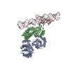
PDB-8huj:
Cryo-EM structure of the J-K-St region of EMCV IRES in complex with eIF4G-HEAT1 and eIF4A
Method: single particle / : Suzuki H, Fujiyoshi Y, Imai S, Shimada I
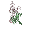
PDB-8j7r:
Cryo-EM structure of the J-K-St region of EMCV IRES in complex with eIF4G-HEAT1 and eIF4A (J-K-St/eIF4G focused)
Method: single particle / : Suzuki H, Fujiyoshi Y, Imai S, Shimada I
PDB-8gcl:
Cryo-EM structure of hAQP2 in DDM
Method: single particle / : Kamegawa A, Suzuki S, Nishikawa K, Numoto N, Suzuki H, Fujiyoshi Y
PDB-8if3:
Structure of human alpha-2/delta-1 with mirogabalin
Method: single particle / : Kozai D, Numoto N, Fujiyoshi Y
PDB-8if4:
Structure of human alpha-2/delta-1 without mirogabalin
Method: single particle / : Kozai D, Numoto N, Fujiyoshi Y
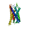
PDB-7wsv:
Cryo-EM structure of the N-terminal deletion mutant of human pannexin-1 in a nanodisc
Method: single particle / : Kuzuya M, Hirano H, Hayashida K, Watanabe M, Kobayashi K, Tani K, Fujiyoshi Y, Oshima A
PDB-7f8j:
Cryo-EM structure of human pannexin-1 in a nanodisc
Method: single particle / : Kuzuya M, Hirano H, Hayashida K, Watanabe M, Kobayashi K, Tani K, Fujiyoshi Y, Oshima A
PDB-7f8n:
Human pannexin-1 showing a conformational change in the N-terminal domain and blocked pore
Method: single particle / : Kuzuya M, Hirano H, Hayashida K, Watanabe M, Kobayashi K, Tani K, Fujiyoshi Y, Oshima A
PDB-7f8o:
Cryo-EM structure of the C-terminal deletion mutant of human PANX1 in a nanodisc
Method: single particle / : Kuzuya M, Hirano H, Hayashida K, Watanabe M, Kobayashi K, Tani K, Fujiyoshi Y, Oshima A
PDB-6kff:
Undocked INX-6 hemichannel in a nanodisc
Method: single particle / : Burendei B, Shinozaki R, Watanabe M, Terada T, Tani K, Fujiyoshi Y, Oshima A
PDB-6kfg:
Undocked INX-6 hemichannel in detergent
Method: single particle / : Burendei B, Shinozaki R, Watanabe M, Terada T, Tani K, Fujiyoshi Y, Oshima A
PDB-6kfh:
Undocked hemichannel of an N-terminal deletion mutant of INX-6 in a nanodisc
Method: single particle / : Burendei B, Shinozaki R, Watanabe M, Terada T, Tani K, Fujiyoshi Y, Oshima A
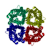
PDB-6acf:
structure of leucine dehydrogenase from Geobacillus stearothermophilus by cryo-EM
Method: single particle / : Yamaguchi H, Kamegawa A, Nakata K, Kashiwagi T, Mizukoshi T, Fujiyoshi Y, Tani K
PDB-6ach:
Structure of NAD+-bound leucine dehydrogenase from Geobacillus stearothermophilus by cryo-EM
Method: single particle / : Yamaguchi H, Kamegawa A, Nakata K, Kashiwagi T, Mizukoshi T, Fujiyoshi Y, Tani K

PDB-5y0b:
PIG GASTRIC H+,K+ - ATPASE IN COMPLEX with BYK99
Method: electron crystallography / : Abe K, Shimokawa J, Natio M, Munson K, Vagin O, Sachs G, Suzuki H, Tani K, Fujiyoshi Y

PDB-5h1q:
C. elegans INX-6 gap junction hemichannel
Method: single particle / : Oshima A, Tani K, Fujiyoshi Y

PDB-5h1r:
C. elegans INX-6 gap junction channel
Method: single particle / : Oshima A, Tani K, Fujiyoshi Y
PDB-4ux1:
Cryo-EM structure of antagonist-bound E2P gastric H,K-ATPase (SCH.E2. AlF)
Method: electron crystallography / : Abe K, Tani K, Fujiyoshi Y
PDB-4ux2:
Cryo-EM structure of antagonist-bound E2P gastric H,K-ATPase (SCH.E2. MgF)
Method: electron crystallography / : Abe K, Tani K, Fujiyoshi Y

PDB-4bgn:
cryo-EM structure of the NavCt voltage-gated sodium channel
Method: electron crystallography / : Tsai CJ, Tani K, Irie K, Hiroaki Y, Shimomura T, Mcmillan DG, Cook GM, Schertler G, Fujiyoshi Y, Li XD

PDB-2yn9:
Cryo-EM structure of gastric H+,K+-ATPase with bound rubidium
Method: electron crystallography / : Abe K, Tani K, Friedrich T, Fujiyoshi Y
PDB-4aq5:
Gating movement in acetylcholine receptor analysed by time-resolved electron cryo-microscopy (closed class)
Method: helical / : Unwin N, Fujiyoshi Y

PDB-4aq9:
Gating movement in acetylcholine receptor analysed by time- resolved electron cryo-microscopy (open class)
Method: helical / : Unwin N, Fujiyoshi Y
PDB-2xzb:
Pig Gastric H,K-ATPase with bound BeF and SCH28080
Method: electron crystallography / : Abe K, Tani K, Fujiyoshi Y

PDB-3iz1:
C-alpha model fitted into the EM structure of Cx26M34A
Method: electron crystallography / : Oshima A, Tani K, Toloue MM, Hiroaki Y, Smock A, Inukai S, Cone A, Nicholson BJ, Sosinsky GE, Fujiyoshi Y

PDB-3iz2:
C-alpha model fitted into the EM structure of Cx26M34Adel2-7
Method: electron crystallography / : Oshima A, Tani K, Toloue MM, Hiroaki Y, Smock A, Inukai S, Cone A, Nicholson BJ, Sosinsky GE, Fujiyoshi Y

PDB-3iyz:
Structure of Aquaporin-4 S180D mutant at 10.0 A resolution from electron micrograph
Method: electron crystallography / : Mitsuma T, Tani K, Hiroaki Y, Kamegawa A, Suzuki H, Hibino H, Kurachi Y, Fujiyoshi Y

PDB-3ixz:
Pig gastric H+/K+-ATPase complexed with aluminium fluoride
Method: electron crystallography / : Abe K, Tani K, Nishizawa T, Fujiyoshi Y
PDB-2zz9:
Structure of aquaporin-4 S180D mutant at 2.8 A resolution by electron crystallography
Method: electron crystallography / : Tani K, Mitsuma T, Hiroaki Y, Kamegawa A, Nishikawa K, Tanimura Y, Fujiyoshi Y

PDB-2d57:
Double layered 2D crystal structure of AQUAPORIN-4 (AQP4M23) at 3.2 a resolution by electron crystallography
Method: electron crystallography / : Hiroaki Y, Tani K, Kamegawa A, Gyobu N, Nishikawa K, Suzuki H, Walz T, Sasaki S, Mitsuoka K, Kimura K, Mizoguchi A, Fujiyoshi Y

PDB-2b6o:
Electron crystallographic structure of lens Aquaporin-0 (AQP0) (lens MIP) at 1.9A resolution, in a closed pore state
Method: electron crystallography / : Gonen T, Cheng Y, Sliz P, Hiroaki Y, Fujiyoshi Y, Harrison SC, Walz T
PDB-1oed:
STRUCTURE OF ACETYLCHOLINE RECEPTOR PORE FROM ELECTRON IMAGES
Method: helical / : Miyazawa A, Fujiyoshi Y, Unwin N
PDB-1fqy:
STRUCTURE OF AQUAPORIN-1 AT 3.8 A RESOLUTION BY ELECTRON CRYSTALLOGRAPHY
Method: electron crystallography / : Murata K, Mitsuoka K, Hirai T, Walz T, Agre P, Heymann JB, Engel A, Fujiyoshi Y
PDB-2at9:
STRUCTURE OF BACTERIORHODOPSIN AT 3.0 ANGSTROM BY ELECTRON CRYSTALLOGRAPHY
Method: electron crystallography / : Mitsuoka K, Hirai T, Murata K, Miyazawa A, Kidera A, Kimura Y, Fujiyoshi Y
PDB-1at9:
STRUCTURE OF BACTERIORHODOPSIN AT 3.0 ANGSTROM DETERMINED BY ELECTRON CRYSTALLOGRAPHY
Method: electron crystallography / : Kimura Y, Vassylyev DG, Miyazawa A, Kidera A, Matsushima M, Mitsuoka K, Murata K, Hirai T, Fujiyoshi Y
 wwPDB to switch to version 3 of the EMDB data model
