 Movie
Movie Controller
 Structure viewers
 About EMN search


-Search query
-Search result
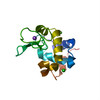
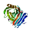
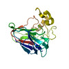
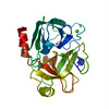
Showing 1 - 50 of 60 items for (author: de & la & cruz & f)

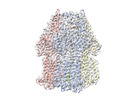
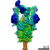
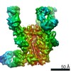
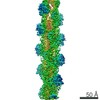
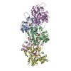
EMDB-41583: 
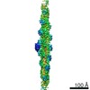
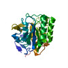
Actin 1 from T. gondii in filaments bound to MgADP
Method: helical / : Hvorecny KL, Sladewski TE, Heaslip AT, Kollman JM

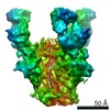
EMDB-41584:
Actin 1 from T. gondii in filaments bound to MgADP and jasplakinolide
Method: helical / : Hvorecny KL, Sladewski TE, Heaslip AT, Kollman JM

PDB-8trm:
Actin 1 from T. gondii in filaments bound to MgADP
Method: helical / : Hvorecny KL, Sladewski TE, Heaslip AT, Kollman JM

PDB-8trn:
Actin 1 from T. gondii in filaments bound to MgADP and jasplakinolide
Method: helical / : Hvorecny KL, Sladewski TE, Heaslip AT, Kollman JM
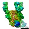
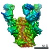
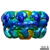
EMDB-17350:
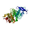
Single particle cryo-EM co-structure of Klebsiella pneumoniae AcrB with the BDM91288 efflux pump inhibitor at 2.97 Angstrom resolution
Method: single particle / : Boernsen C, Mueller RT, Pos KM, Frangakis AS
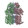
PDB-8p1i:
Single particle cryo-EM co-structure of Klebsiella pneumoniae AcrB with the BDM91288 efflux pump inhibitor at 2.97 Angstrom resolution
Method: single particle / : Boernsen C, Mueller RT, Pos KM, Frangakis AS

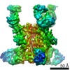
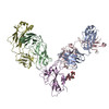
EMDB-15213:
Cryo-EM density map of Tn4430 TnpA hyperactive mutant (TnpA3X) in complex with IR48 substrate.
Method: single particle / : Shkumatov AV, Liu Y, Efremov RG

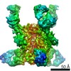
EMDB-15218:
Medium resolution cryo-EM density map of Tn4430 TnpA transposase from Tn3 family in apo state
Method: single particle / : Shkumatov AV, Liu Y, Efremov RG
EMDB-25132:
Androgen receptor bound to DNA - Entrenched state
Method: single particle / : Wasmuth EV, Vanden Broeck A, Klinge S, Sawyers CL

EMDB-25133:
Androgen receptor bound to DNA - Splayed state
Method: single particle / : Wasmuth EV, Vanden Broeck A, Klinge S, Sawyers CL
EMDB-25134:
Androgen receptor bound to DNA - Divorced state
Method: single particle / : Wasmuth EV, Vanden Broeck A, Klinge S, Sawyers CL
EMDB-25105:
Structure of SARS-CoV S protein in complex with Receptor Binding Domain antibody DH1047
Method: single particle / : Gobeil S, Acharya P

PDB-7sg4:
Structure of SARS-CoV S protein in complex with Receptor Binding Domain antibody DH1047
Method: single particle / : Gobeil S, Acharya P
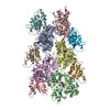
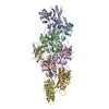
EMDB-22170:
BG505 SOSIPv5.2 in complex with PGT122 and two RM20E1 Fabs
Method: single particle / : Cottrell CA, Ward AB
EMDB-22171:
BG505 SOSIPv5.2 in complex with PGT122 and three RM20E1 Fabs
Method: single particle / : Cottrell CA, Ward AB
EMDB-22172:
C3 symmetric BG505 SOSIPv5.2 in complex with PGT122 and three RM20E1 Fabs
Method: single particle / : Cottrell CA, Ward AB
EMDB-22173:
BG505 SOSIPv5.2 in complex with PGT122 and one RM20E1 Fab
Method: single particle / : Cottrell CA, Ward AB
EMDB-22178:
BG505 SOSIPv5.2 in complex with PGT122 and no RM20E1 Fabs
Method: single particle / : Cottrell CA, Ward AB
EMDB-22179:
C3 symmetric BG505 SOSIPv5.2 in complex with PGT122 and no RM20E1 Fabs
Method: single particle / : Cottrell CA, Ward AB

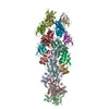
EMDB-22137:
Complex of SARS-CoV-2 receptor binding domain with the Fab fragments of two neutralizing antibodies
Method: single particle / : Franklin MC, Saotome K, Romero Hernandez A, Zhou Y
PDB-6xdg:
Complex of SARS-CoV-2 receptor binding domain with the Fab fragments of two neutralizing antibodies
Method: single particle / : Franklin MC, Saotome K, Romero Hernandez A, Zhou Y
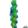
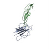
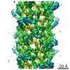
EMDB-20711:
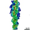
Cryo-EM structure of cofilactin from partially cofilin-decorated actin filaments.
Method: helical / : Huehn AR, Bibeau JP, Schramm AC, Cao W, De La Cruz EM, Sindelar CV
PDB-6vao:
Human cofilin-1 decorated actin filament
Method: helical / : Huehn AR, Bibeau JP, Schramm AC, Cao W, De La Cruz EM, Sindelar CV
EMDB-20719:
Bare actin from partially cofilin-decorated actin filaments
Method: helical / : Huehn AR, Bibeau JP, Schramm AC, Cao W, De La Cruz EM, Sindelar CV
EMDB-20721:
Isolated cofilin bound to an actin filament
Method: single particle / : Huehn AR, Bibeau JP, Schramm AC, Cao W, De La Cruz EM, Sindelar CV
EMDB-20724:
Isolated S3D-cofilin bound to an actin filament
Method: single particle / : Huehn AR, Bibeau JP, Schramm AC, Cao W, De La Cruz EM, Sindelar CV
EMDB-20726:
Barbed end side of a cofilactin cluster
Method: single particle / : Huehn AR, Bibeau JP, Schramm AC, Cao W, De La Cruz EM, Sindelar CV

PDB-6uby:
Isolated cofilin bound to an actin filament
Method: single particle / : Huehn AR, Bibeau JP, Schramm AC, Cao W, De La Cruz EM, Sindelar CV
PDB-6uc0:
Isolated S3D-cofilin bound to an actin filament
Method: single particle / : Huehn AR, Bibeau JP, Schramm AC, Cao W, De La Cruz EM, Sindelar CV
PDB-6uc4:
Barbed end side of a cofilactin cluster
Method: single particle / : Huehn AR, Bibeau JP, Schramm AC, Cao W, De La Cruz EM, Sindelar CV
PDB-6vau:
Bare actin filament from a partially cofilin-decorated sample
Method: helical / : Huehn AR, Bibeau JP, Schramm AC, Cao W, De La Cruz EM, Sindelar CV
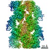
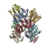
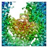
EMDB-7321:
Integrative Structure and Functional Anatomy of a Nuclear Pore Complex
Method: subtomogram averaging / : Kim SJ, Fernandez-Martinez J, Nudelman I, Shi Y, Zhang W, Ludtke SJ, Akey CW, Chait BT, Sali A, Rout MP
PDB-5ty4:
MicroED structure of a complex between monomeric TGF-b and its receptor, TbRII, at 2.9 A resolution
Method: electron crystallography / : Weiss SC, de la Cruz MJ, Hattne J, Shi D, Reyes FE, Callero G, Gonen T
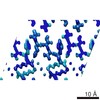
PDB-5k7n:
MicroED structure of tau VQIVYK peptide at 1.1 A resolution
Method: electron crystallography / : de la Cruz MJ, Hattne J, Shi D, Seidler P, Rodriguez J, Reyes FE, Sawaya MR, Cascio D, Eisenberg D, Gonen T
PDB-5k7o:
MicroED structure of lysozyme at 1.8 A resolution
Method: electron crystallography / : de la Cruz MJ, Hattne J, Shi D, Seidler P, Rodriguez J, Reyes FE, Sawaya MR, Cascio D, Eisenberg D, Gonen T
PDB-5k7p:
MicroED structure of xylanase at 2.3 A resolution
Method: electron crystallography / : de la Cruz MJ, Hattne J, Shi D, Seidler P, Rodriguez J, Reyes FE, Sawaya MR, Cascio D, Eisenberg D, Gonen T
PDB-5k7q:
MicroED structure of thaumatin at 2.5 A resolution
Method: electron crystallography / : de la Cruz MJ, Hattne J, Shi D, Seidler P, Rodriguez J, Reyes FE, Sawaya MR, Cascio D, Eisenberg D, Gonen T
PDB-5k7r:
MicroED structure of trypsin at 1.7 A resolution
Method: electron crystallography / : de la Cruz MJ, Hattne J, Shi D, Seidler P, Rodriguez J, Reyes FE, Sawaya MR, Cascio D, Eisenberg D, Gonen T
PDB-5k7s:
MicroED structure of proteinase K at 1.6 A resolution
Method: electron crystallography / : de la Cruz MJ, Hattne J, Shi D, Seidler P, Rodriguez J, Reyes FE, Sawaya MR, Cascio D, Eisenberg D, Gonen T
PDB-5k7t:
MicroED structure of thermolysin at 2.5 A resolution
Method: electron crystallography / : de la Cruz MJ, Hattne J, Shi D, Seidler P, Rodriguez J, Reyes FE, Sawaya MR, Cascio D, Eisenberg D, Gonen T
EMDB-8300:
Cryo-EM structure of Casp-8 tDED filament (CASP target)
Method: helical / : Fu TM, Li Y, Wu H
EMDB-8216:
MicroED structure of tau VQIVYK peptide at 1.1 A resolution
Method: electron crystallography / : de la Cruz MJ, Hattne J
EMDB-8217:
MicroED structure of lysozyme at 1.8 A resolution
Method: electron crystallography / : de la Cruz MJ, Hattne J, Shi D, Seidler P, Rodriguez J, Reyes FE, Sawaya MR, Cascio D, Eisenberg D, Gonen T
EMDB-8218:
MicroED structure of xylanase at 2.3 A resolution
Method: electron crystallography / : de la Cruz MJ, Hattne J
EMDB-8219:
MicroED structure of thaumatin at 2.5 A resolution
Method: electron crystallography / : de la Cruz MJ, Hattne J, Shi D, Seidler P, Rodriguez J, Reyes FE, Sawaya MR, Cascio D, Eisenberg D, Gonen T

EMDB-8220:
MicroED structure of trypsin at 1.7 A resolution
Method: electron crystallography / : de la Cruz MJ, Hattne J, Shi D, Seidler P, Rodriguez J, Reyes FE, Sawaya MR, Cascio D, Eisenberg D, Gonen T
EMDB-8221:
MicroED structure of proteinase K at 1.6 A resolution
Method: electron crystallography / : de la Cruz MJ, Hattne J, Shi D, Seidler P, Rodriguez J, Reyes FE, Sawaya MR, Cascio D, Eisenberg D, Gonen T
EMDB-8222:
MicroED structure of thermolysin at 2.5 A resolution
Method: electron crystallography / : de la Cruz MJ, Hattne J
EMDB-8472:
MicroED structure of a complex between monomeric TGF-b and its receptor, TbRII, at 2.9 A resolution
Method: electron crystallography / : Weiss SC, de la Cruz MJ
Pages:
 wwPDB to switch to version 3 of the EMDB data model
