 Movie
Movie Controller
 Structure viewers
 About EMN search


-Search query
-Search result
Showing 1 - 50 of 67 items for (author: de & haas & f)
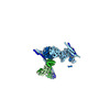
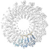
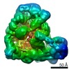
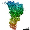
EMDB-44479: 
Cryo-EM structure of synthetic claudin-4 complex with Clostridium perfringens enterotoxin C-terminal domain, sFab COP-2, and Nanobody
Method: single particle / : Vecchio AJ

PDB-9bei:
Cryo-EM structure of synthetic claudin-4 complex with Clostridium perfringens enterotoxin C-terminal domain, sFab COP-2, and Nanobody
Method: single particle / : Vecchio AJ

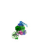
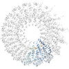
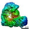
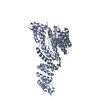
EMDB-16730:
Cryo-EM structure of complement C5 in complex with nanobodies UNbC5-1 and UNbC5-2
Method: single particle / : De la O Becerra KI, Gros P

PDB-8cml:
Cryo-EM structure of complement C5 in complex with nanobodies UNbC5-1 and UNbC5-2
Method: single particle / : De la O Becerra KI, Gros P
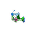
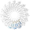
EMDB-14808:
Structure of the human TREX core THO-UAP56 complex (map D)
Method: single particle / : Pacheco-Fiallos FB, Vorlaender MK, Plaschka C
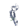
EMDB-17215:
Composite map of the human TREX core THO-UAP56 complex
Method: single particle / : Pacheco-Fiallos FB, Vorlaender MK, Plaschka C

PDB-7znl:
Structure of the human TREX core THO-UAP56 complex
Method: single particle / : Pacheco-Fiallos FB, Vorlaender MK, Plaschka C
EMDB-14804:
Structure of an endogenous human TREX complex bound to mRNA, composite map
Method: single particle / : Pacheco-Fiallos FB, Vorlaender MK, Plaschka C

EMDB-16753:
Tomogram of purified human TREX-mRNPs
Method: electron tomography / : Pacheco-Fiallos FB, Vorlaender MK, Plaschka C

PDB-7znk:
Structure of an endogenous human TREX complex bound to mRNA
Method: single particle / : Pacheco-Fiallos FB, Vorlaender MK, Plaschka C
EMDB-14805:
Structure of an endogenous human TREX complex bound to mRNA, map A
Method: single particle / : Pacheco-Fiallos FB, Vorlaender MK, Plaschka C
EMDB-14806:
Structure of an endogenous human TREX complex bound to mRNA, map B
Method: single particle / : Pacheco-Fiallos FB, Vorlaender MK, Plaschka C
EMDB-14807:
Structure of an endogenous human TREX complex bound to mRNA, map C
Method: single particle / : Pacheco-Fiallos FB, Vorlaender MK, Plaschka C
EMDB-14809:
Structure of the human TREX core THO-UAP56 complex (map E)
Method: single particle / : Pacheco-Fiallos FB, Vorlaender MK, Plaschka C

EMDB-16633:
Structure of an ALYREF-exon junction complex hexamer, obtained from TREX-EJC-RNA sample
Method: single particle / : Pacheco-Fiallos FB, Vorlaender MK, Plaschka C
EMDB-14803:
Structure of an ALYREF-exon junction complex hexamer
Method: single particle / : Pacheco-Fiallos FB, Vorlaender MK, Plaschka C

PDB-7znj:
Structure of an ALYREF-exon junction complex hexamer
Method: single particle / : Pacheco-Fiallos FB, Vorlaender MK, Plaschka C
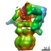
EMDB-14407:
Single particle structure of keyhole limpet hemocyanin obtained via iDPC scanning transmission electron microscopy
Method: single particle / : Mann D, Lazic I, Wirix M, de Haas F, Sachse C
EMDB-13778:
cryo iDPC-STEM structure recorded with CSA 2.0
Method: helical / : Sachse C, Leidl ML
EMDB-13779:
cryo iDPC-STEM structure recorded with CSA 3.0
Method: helical / : Sachse C, Leidl ML
EMDB-13780:
cryo iDPC-STEM structure recorded with CSA 3.5
Method: helical / : Sachse C, Leidl ML
EMDB-13781:
cryo iDPC-STEM structure recorded with CSA 4.0
Method: helical / : Sachse C, Leidl ML
EMDB-13782:
cryo iDPC-STEM structure recorded with CSA 4.5
Method: helical / : Sachse C, Leidl ML
EMDB-32216:
26S proteasome from the cell with TDP-25 inclusion
Method: subtomogram averaging / : Guo Q
EMDB-32217:
tomogram of a rat primary neuron harboring TDP-25 inclusion
Method: electron tomography / : Guo Q
EMDB-25026:
Anaphase Promoting Complex delta APC7
Method: single particle / : Ferguson CJ, Brown NG, Prabu JR, Watson ER, Schulman BA, Bonni A
EMDB-25027:
Anaphase Promoting Complex delta APC7 + UBE2C,substrate,UbV,CDH1
Method: single particle / : Ferguson CJ, Brown NG, Prabu JR, Watson ER, Schulman BA, Bonni A
EMDB-22829:
Human Tom70 in complex with SARS CoV2 Orf9b
Method: single particle / : QCRG Structural Biology Consortium
PDB-7kdt:
Human Tom70 in complex with SARS CoV2 Orf9b
Method: single particle / : QCRG Structural Biology Consortium

PDB-6zet:
Crystal structure of proteinase K nanocrystals by electron diffraction with a 20 micrometre C2 condenser aperture
Method: electron crystallography / : Evans G, Zhang P, Beale EV, Waterman DG

PDB-6zeu:
Crystal structure of proteinase K lamella by electron diffraction with a 50 micrometre C2 condenser aperture
Method: electron crystallography / : Evans G, Zhang P, Beale EV, Waterman DG

PDB-6zev:
Crystal structure of proteinase K lamellae by electron diffraction with a 20 micrometre C2 condenser aperture
Method: electron crystallography / : Evans G, Zhang P, Beale EV, Waterman DG
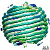
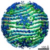
EMDB-10289:
Bacteriophage phi6 dsRNA genome, layer 1, conformation pseudo C2
Method: single particle / : Ilca SL, Huiskonen JT
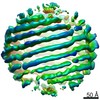
EMDB-10075:
Bacteriophage phi6 dsRNA genome, layer 1, conformation pseudo D3
Method: single particle / : Ilca SL, Huiskonen JT
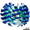
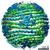
EMDB-0294:
Bacteriophage phi6 dsRNA genome, layer 2
Method: single particle / : Ilca SL, Huiskonen JT
EMDB-0295:
Bacteriophage phi6 dsRNA genome, layer 3
Method: single particle / : Ilca SL, Huiskonen JT
EMDB-0296:
Bacteriophage phi6 dsRNA genome, layers 4 and 5
Method: single particle / : Ilca SL, Huiskonen JT
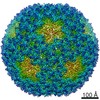
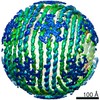
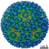
EMDB-0299:
Bacteriophage phi6 nucleocapsid reconstructed with icosahedral symmetry
Method: single particle / : Ilca SL, Huiskonen JT
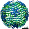
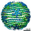
EMDB-0300:
Reconstruction of dsRNA bacteriophage phi6 nucleocapsid with D3 symmetry
Method: single particle / : Ilca SL, Huiskonen JT
EMDB-0302:
Bacteriophage phi6 dsRNA genome, layer 1, conformation pseudo D3'
Method: single particle / : Ilca SL, Huiskonen JT
EMDB-0304:
Bacteriophage phi6 dsRNA genome, layer 1, conformation pseudo D3', sub-conformation 1
Method: single particle / : Ilca SL, Huiskonen JT
EMDB-0305:
Bacteriophage phi6 dsRNA genome, layer 1, conformation pseudo D3', sub-conformation 2
Method: single particle / : Ilca SL, Huiskonen JT
EMDB-0306:
Bacteriophage phi6 dsRNA genome, layer 1, conformation pseudo D3', sub-conformation 3
Method: single particle / : Ilca SL, Huiskonen JT

PDB-6hy0:
Atomic models of P1, P4 C-terminal fragment and P8 fitted in the bacteriophage phi6 nucleocapsid reconstructed with icosahedral symmetry
Method: single particle / : El Omari K, Ilca SL, Stuart DI, Huiskonen JT
EMDB-9779:
Reconstruction of HRPV6 VP5 spike
Method: subtomogram averaging / : Li S, Huiskonen JT
Pages:
 wwPDB to switch to version 3 of the EMDB data model
