 ムービー
ムービー コントローラー
 構造ビューア
 EMN検索について


-検索条件
-検索結果
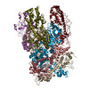
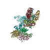
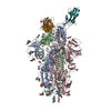
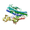
検索 (著者・登録者: d. & clare)の結果54件中、1から50件目までを表示しています

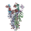
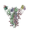
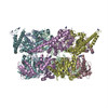
PDB-7r5o: 
1.58 A STRUCTURE OF HUMAN APOFERRITIN OBTAINED FROM TITAN KRIOS 2 AT eBIC, DLS UNDER COMMISSIONING SESSION CM26464-2
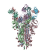
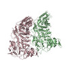
PDB-7q6e:
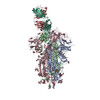
Beta049 fab in complex with SARS-CoV2 beta-Spike glycoprotein, The Beta mAb response underscores the antigenic distance to other SARS-CoV-2 variants
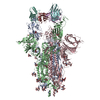
PDB-7q9f:
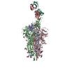
Beta-50 fab in complex with SARS-CoV-2 beta-Spike glycoprotein
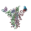
PDB-7q9g:
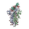
COVOX-222 fab in complex with SARS-CoV-2 beta-Spike glycoprotein
PDB-7q9i:
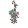
Beta-43 fab in complex with SARS-CoV-2 beta-Spike glycoprotein
PDB-7q9j:
Beta-26 fab in complex with SARS-CoV-2 beta-Spike glycoprotein
PDB-7q9k:
Beta-32 fab in complex with SARS-CoV-2 beta-Spike glycoprotein
PDB-7q9m:
Beta-53 fab in complex with SARS-CoV-2 beta-Spike glycoprotein
PDB-7q9p:
Beta-06 fab in complex with SARS-CoV-2 beta-Spike glycoprotein
PDB-7nd3:
EM structure of SARS-CoV-2 Spike glycoprotein in complex with COVOX-40 Fab
PDB-7nd4:
EM structure of SARS-CoV-2 Spike glycoprotein in complex with COVOX-88 Fab

PDB-7nd5:
EM structure of SARS-CoV-2 Spike glycoprotein in complex with COVOX-150 Fab
PDB-7nd6:
EM structure of SARS-CoV-2 Spike glycoprotein in complex with COVOX-40 Fab
PDB-7nd7:
EM structure of SARS-CoV-2 Spike glycoprotein in complex with COVOX-316 Fab
PDB-7nd8:
EM structure of SARS-CoV-2 Spike glycoprotein in complex with COVOX-384 Fab
PDB-7nd9:
EM structure of SARS-CoV-2 Spike glycoprotein (one RBD up) in complex with COVOX-253H55L Fab
PDB-7nda:
EM structure of SARS-CoV-2 Spike glycoprotein (all RBD down) in complex with COVOX-253H55L Fab
PDB-7ndb:
EM structure of SARS-CoV-2 Spike glycoprotein in complex with COVOX-253H165L Fab
PDB-7ndc:
EM structure of SARS-CoV-2 Spike glycoprotein (all RBD down) in complex with COVOX-159

PDB-7ndd:
EM structure of SARS-CoV-2 Spike glycoprotein (one RBD up) in complex with COVOX-159
PDB-7b3y:
Structure of a nanoparticle for a COVID-19 vaccine candidate
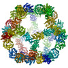
PDB-6xf7:
SLP
PDB-6xf8:
DLP 5 fold

PDB-6zts:
Assembly intermediates of orthoreovirus captured in the cell
PDB-6zty:
Assembly intermediates of orthoreovirus captured in the cell
PDB-6ztz:
Assembly intermediates of orthoreovirus captured in the cell

PDB-6zhd:
H11-H4 bound to Spike
PDB-6z43:
Cryo-EM Structure of SARS-CoV-2 Spike : H11-D4 Nanobody Complex

PDB-6vwl:
70S ribosome bound to HIV frameshifting stem-loop (FSS) and P/E tRNA (rotated conformation)

PDB-6vwm:
70S ribosome bound to HIV frameshifting stem-loop (FSS) and P-site tRNA (non-rotated conformation, Structure I)

PDB-6vwn:
70S ribosome bound to HIV frameshifting stem-loop (FSS) and P-site tRNA (non-rotated conformation, Structure II)

PDB-6i3n:
Helical MyD88 death domain filament
PDB-6j7v:
Structure of HRPV6 VP5 fitted in the cryoEM density of the spike

PDB-6ne0:
Structure of double-stranded target DNA engaged Csy complex from Pseudomonas aeruginosa (PA-14)

PDB-5nbz:
Wzz dodecamer fitted by MDFF to the Wzz experimental map from cryo-EM
PDB-5a79:
Novel inter-subunit contacts in Barley Stripe Mosaic Virus revealed by cryo-EM
PDB-5a7a:
Novel inter-subunit contacts in Barley Stripe Mosaic Virus revealed by cryo-EM
PDB-4d2q:
Negative-stain electron microscopy of E. coli ClpB mutant E432A (BAP form bound to ClpP)
PDB-4d2u:
Negative-stain electron microscopy of E. coli ClpB (BAP form bound to ClpP)
PDB-4d2x:
Negative-stain electron microscopy of E. coli ClpB of Y503D hyperactive mutant (BAP form bound to ClpP)
PDB-2m5k:
Atomic-resolution structure of a doublet cross-beta amyloid fibril
PDB-2m5m:
Atomic-resolution structure of a triplet cross-beta amyloid fibril
PDB-3zpk:
Atomic-resolution structure of a quadruplet cross-beta amyloid fibril.
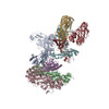
PDB-4aaq:
ATP-triggered molecular mechanics of the chaperonin GroEL
PDB-4aar:
ATP-triggered molecular mechanics of the chaperonin GroEL
PDB-4aas:
ATP-triggered molecular mechanics of the chaperonin GroEL
PDB-4aau:
ATP-triggered molecular mechanics of the chaperonin GroEL
PDB-4ab2:
ATP-triggered molecular mechanics of the chaperonin GroEL
PDB-4ab3:
ATP-triggered molecular mechanics of the chaperonin GroEL
PDB-2xrp:
Human Doublecortin N-DC Repeat (1MJD) and Mammalian Tubulin (1JFF and 3HKE) Docked into the 8-Angstrom Cryo-EM Map of Doublecortin- Stabilised Microtubules
ページ:
 wwPDBはEMDBデータモデルのバージョン3へ移行します
