 ムービー
ムービー コントローラー
 構造ビューア
 EMN検索について


-検索条件
-検索結果
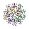
検索 (著者・登録者: bharat, & t.a.m)の結果全22件を表示しています
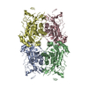
PDB-8q7y: 
ESIBD structure of beta-galactosidase
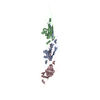
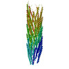
PDB-8ch5:
Cryo-EM structure of the fd bacteriophage capsid major coat protein pVIII
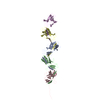
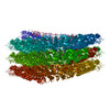
PDB-8cio:
Cryo-EM structure of the CupE pilus from Pseudomonas aeruginosa
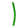
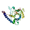
PDB-8aur:
Cryo-EM structure of a TasA fibre

PDB-8ae1:
Structure of trimeric SlpA outer membrane protein

PDB-7ptp:
In-situ structure of pentameric S-layer protein

PDB-7ptr:
Structure of hexameric S-layer protein from Haloferax volcanii archaea

PDB-7ptt:
In-situ structure of hexameric S-layer protein
PDB-7ptu:
Structure of pentameric S-layer protein from Halofaerax volcanii

PDB-7peo:
Structure of the Caulobacter crescentus S-layer protein RsaA N-terminal domain bound to LPS and soaked with Holmium

PDB-6z7p:
Composite model of the Caulobacter crescentus S-layer bound to the O-antigen of lipopolysaccharide
PDB-6tup:
Cryo-EM structure of Pf4 bacteriophage coat protein with single-stranded DNA

PDB-6tuq:
Cryo-EM structure of Pf4 bacteriophage coat protein without ssDNA

PDB-6t72:
Structure of the RsaA N-terminal domain bound to LPS

PDB-5o09:
BtubABC mini microtubule

PDB-5aey:
actin-like ParM protein bound to AMPPNP

PDB-5ai7:
ParM doublet model
PDB-4udv:
Cryo-EM structure of TMV at 3.35 A resolution

PDB-4d1k:
Cryo-electron microscopy of tubular arrays of HIV-1 Gag resolves structures essential for immature virus assembly.
PDB-4blf:
Variable internal flexibility characterizes the helical capsid formed by Agrobacterium VirE2 protein on single-stranded DNA.
PDB-4ard:
Structure of the immature retroviral capsid at 8A resolution by cryo- electron microscopy

PDB-4arg:
Structure of the immature retroviral capsid at 8A resolution by cryo- electron microscopy
 wwPDBはEMDBデータモデルのバージョン3へ移行します
