 ムービー
ムービー コントローラー
 構造ビューア
 EMN検索について


-検索条件
-検索結果
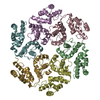
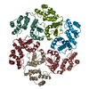
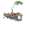
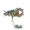
検索 (著者・登録者: a. & sen)の結果772件中、1から50件目までを表示しています
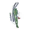
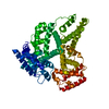
PDB-8kab: 
Mycobacterium smegmatis 50S ribosomal subunit-HflX complex
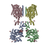
PDB-8xz3:
Mycobacterium smegmatis 50S ribosomal subunit with Erythromycin
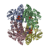
PDB-8qb7:
Pil1 in native eisosome lattice bound to plasma membrane microdomain
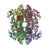
PDB-8qb8:
Lsp1 in native eisosome lattice bound to plasma membrane microdomain
PDB-8qb9:
Helical reconstruction of yeast eisosome protein Pil1 bound to membrane composed of lipid mixture -PIP2/+sterol (DOPC, DOPE, DOPS, cholesterol 30:20:20:30)
PDB-8qbb:
Helical reconstruction of yeast eisosome protein Pil1 bound to membrane composed of lipid mixture +PIP2/-sterol (DOPC, DOPE, DOPS, PI(4,5)P2 50:20:20:10)
PDB-8qbd:
Helical reconstruction of yeast eisosome protein Pil1 bound to membrane composed of lipid mixture +PIP2/+sterol (DOPC, DOPE, DOPS, cholesterol, PI(4,5)P2 35:20:20:15:10)
PDB-8qbe:
Compact state - Pil1 in native eisosome lattice bound to plasma membrane microdomain
PDB-8qbf:
Compact state - Pil1 dimer with lipid headgroups fitted in native eisosome lattice bound to plasma membrane microdomain
PDB-8qbg:
Stretched state - Pil1 in native eisosome lattice bound to plasma membrane microdomain
PDB-9euo:
Outward-open structure of Drosophila dopamine transporter bound to an atypical non-competitive inhibitor
PDB-9eup:
Inhibitor-free outward-open structure of Drosophila dopamine transporter
PDB-8ozh:
In situ cryoEM structure of Prototype Foamy Virus Env trimer
PDB-8ozj:
In situ cryoEM structure of Prototype Foamy Virus Env dimer of trimers
PDB-8ozk:
In situ cryoEM structure of the Prototype Foamy Virus capsid, icosahedral map
PDB-8ozl:
In situ cryoEM structure of the Prototype Foamy Virus capsid, pentamer localised reconstruction
PDB-8ozm:
In situ cryoEM structure of the Prototype Foamy Virus capsid, hexamer 1 localised reconstruction
PDB-8ozn:
In situ cryoEM structure of the Prototype Foamy Virus capsid, hexamer 2 localised reconstruction
PDB-8ozp:
In situ subtomogram average of Prototype Foamy Virus Env pentamer of trimers
PDB-8ozq:
In situ subtomogram average of Prototype Foamy Virus Env hexamer of trimers

PDB-9eo4:
Outward-open structure of human dopamine transporter bound to cocaine

PDB-8vqy:
Human GABAA receptor alpha1-beta2-gamma2 subtype in complex with GABA plus methaqualone

PDB-8vrn:
Human GABAA receptor alpha1-beta2-gamma2 subtype in complex with GABA plus PPTQ

PDB-8vac:
Cryogenic electron microscopy structure of human serum albumin in complex with teniposide

PDB-8vae:
Cryogenic electron microscopy structure of human serum albumin in complex with salicylic acid
PDB-8vaf:
Cryogenic electron microscopy structure of apo human serum albumin

PDB-9erx:
Structural basis of D9-THC analog activity at the Cannabinoid 1 receptor

PDB-8utn:
KIF1A[1-393] AMP-PNP bound two-heads-bound state in complex with a microtubule (class T23L1)

PDB-8uto:
KIF1A[1-393] AMP-PNP bound two-heads-bound state in complex with a microtubule - class T2L1

PDB-8utp:
KIF1A[1-393] - AMP-PNP two-heads-bound state in complex with a microtubule - class T3L1

PDB-8utq:
KIF1A[1-393] AMP-PNP bound one-head-bound state in complex with a microtubule - class T1L02*

PDB-8utr:
KIF1A[1-393] ADP bound in complex with a microtubule

PDB-8uts:
KIF1A[1-393] APO in complex with a microtubule

PDB-8utt:
KIF1A[1-393] P305L mutant AMP-PNP bound two-heads-bound state in complex with a microtubule

PDB-8utu:
KIF1A[1-393] P305L mutant AMP-PNP bound one and two heads bound states merged, in complex with a microtubule

PDB-8utv:
KIF1A[1-393] P305L mutant ADP bound in complex with a microtubule

PDB-8utw:
KIF1A[1-393] P305L mutant APO in complex with a microtubule

PDB-8uty:
KIF1A[1-393] P364L mutant AMP-PNP bound two-heads-bound state in complex with a microtubule

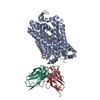
PDB-8p2w:
Structure of human SIT1 (focussed map / refinement)

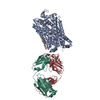
PDB-8p2x:
Structure of human SIT1:ACE2 complex (open PD conformation)

PDB-8p2y:
Structure of human SIT1:ACE2 complex (closed PD conformation)

PDB-8p2z:
Structure of human SIT1 bound to L-pipecolate (focussed map / refinement)
PDB-8p30:
Structure of human SIT1:ACE2 complex (open PD conformation) bound to L-pipecolate

PDB-8p31:
Structure of human SIT1:ACE2 complex (closed PD conformation) bound to L-pipecolate

PDB-8qbk:
Retron-Eco1 filament with ADP-ribosylated Effector (local map with 1 segment)

PDB-8qbl:
Retron-Eco1 filament with inactive effector (E106A, 2 segments)

PDB-8qbm:
Retron-Eco1 filament with ADP-ribosylated Effector (full map with 2 segments)
PDB-8qxj:
Cryo-EM structure of tetrameric human SAMHD1 with dApNHpp
PDB-8qxk:
Cryo-EM structure of tetrameric human SAMHD1 State I - Tense
PDB-8qxl:
Cryo-EM structure of tetrameric human SAMHD1 State II - Hemi-relaxed
ページ:
 wwPDBはEMDBデータモデルのバージョン3へ移行します
