 Movie
Movie Controller


+ Open data

- Basic information
| Entry | Database: PDB / ID: 5k47 | |||||||||
|---|---|---|---|---|---|---|---|---|---|---|
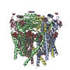
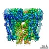
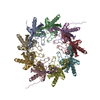
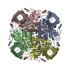
| Title | CryoEM structure of the human Polycystin-2/PKD2 TRP channel | |||||||||
 Components Components | Polycystin-2 Polycystin 2 Polycystin 2 | |||||||||
 Keywords Keywords | TRANSPORT PROTEIN / ion channel / transient receptor potential channel / polycystic kidney disease / Structural Genomics / Structural Genomics Consortium / SGC | |||||||||
| Function / homology |  Function and homology information Function and homology informationdetection of nodal flow / metanephric smooth muscle tissue development / metanephric cortex development / metanephric cortical collecting duct development / metanephric distal tubule development / polycystin complex / mesonephric tubule development / mesonephric duct development / : / metanephric part of ureteric bud development ...detection of nodal flow / metanephric smooth muscle tissue development / metanephric cortex development / metanephric cortical collecting duct development / metanephric distal tubule development / polycystin complex / mesonephric tubule development / mesonephric duct development / : / metanephric part of ureteric bud development / determination of liver left/right asymmetry / renal tubule morphogenesis / metanephric ascending thin limb development / HLH domain binding / basal cortex / metanephric mesenchyme development / metanephric S-shaped body morphogenesis / renal artery morphogenesis / positive regulation of inositol 1,4,5-trisphosphate-sensitive calcium-release channel activity / migrasome / cilium organization / VxPx cargo-targeting to cilium / detection of mechanical stimulus / regulation of calcium ion import / cation channel complex / calcium-induced calcium release activity / muscle alpha-actinin binding / placenta blood vessel development / voltage-gated monoatomic ion channel activity / cellular response to hydrostatic pressure / outward rectifier potassium channel activity / voltage-gated monoatomic cation channel activity / non-motile cilium / cellular response to fluid shear stress / cellular response to osmotic stress / voltage-gated sodium channel activity / transcription regulator inhibitor activity / actinin binding / motile cilium / inorganic cation transmembrane transport / determination of left/right symmetry / neural tube development / aorta development / protein heterotetramerization / ciliary membrane / branching involved in ureteric bud morphogenesis / negative regulation of G1/S transition of mitotic cell cycle / spinal cord development / heart looping / cytoplasmic side of endoplasmic reticulum membrane / voltage-gated potassium channel activity / cell surface receptor signaling pathway via JAK-STAT / potassium channel activity / centrosome duplication / sodium ion transmembrane transport / negative regulation of ryanodine-sensitive calcium-release channel activity / voltage-gated calcium channel activity / embryonic placenta development / monoatomic cation channel activity / cellular response to cAMP / release of sequestered calcium ion into cytosol / potassium ion transmembrane transport / cellular response to calcium ion / cytoskeletal protein binding / basal plasma membrane / ciliary basal body / liver development / establishment of localization in cell / lumenal side of endoplasmic reticulum membrane / calcium ion transmembrane transport / protein tetramerization / phosphoprotein binding / cytoplasmic vesicle membrane / cilium / intracellular calcium ion homeostasis / mitotic spindle / Wnt signaling pathway / cellular response to reactive oxygen species / positive regulation of nitric oxide biosynthetic process / calcium ion transport / cell-cell junction / lamellipodium / regulation of cell population proliferation / heart development / ATPase binding / positive regulation of cytosolic calcium ion concentration / protein homotetramerization / basolateral plasma membrane / transmembrane transporter binding / regulation of cell cycle / negative regulation of cell population proliferation / signaling receptor binding / calcium ion binding / endoplasmic reticulum membrane / positive regulation of gene expression / Golgi apparatus / endoplasmic reticulum / protein homodimerization activity / positive regulation of transcription by RNA polymerase II / extracellular exosomeSimilarity search - Function | |||||||||
| Biological species |  Homo sapiens (human) Homo sapiens (human) | |||||||||
| Method | ELECTRON MICROSCOPY / single particle reconstruction / cryo EM / Resolution: 4.22 Å | |||||||||
 Authors Authors | Pike, A.C.W. / Grieben, M. / Shintre, C.A. / Tessitore, A. / Shrestha, L. / Mukhopadhyay, S. / Mahajan, P. / Chalk, R. / Burgess-Brown, N.A. / Huiskonen, J.T. ...Pike, A.C.W. / Grieben, M. / Shintre, C.A. / Tessitore, A. / Shrestha, L. / Mukhopadhyay, S. / Mahajan, P. / Chalk, R. / Burgess-Brown, N.A. / Huiskonen, J.T. / Arrowsmith, C.H. / Edwards, A.M. / Bountra, C. / Carpenter, E.P. / Structural Genomics Consortium (SGC) | |||||||||
| Funding support |  United Kingdom, 1items United Kingdom, 1items
| |||||||||
 Citation Citation | Journal: Nat Struct Mol Biol / Year: 2017 Title: Structure of the polycystic kidney disease TRP channel Polycystin-2 (PC2). Authors: Mariana Grieben / Ashley C W Pike / Chitra A Shintre / Elisa Venturi / Sam El-Ajouz / Annamaria Tessitore / Leela Shrestha / Shubhashish Mukhopadhyay / Pravin Mahajan / Rod Chalk / Nicola A ...Authors: Mariana Grieben / Ashley C W Pike / Chitra A Shintre / Elisa Venturi / Sam El-Ajouz / Annamaria Tessitore / Leela Shrestha / Shubhashish Mukhopadhyay / Pravin Mahajan / Rod Chalk / Nicola A Burgess-Brown / Rebecca Sitsapesan / Juha T Huiskonen / Elisabeth P Carpenter / Abstract: Mutations in either polycystin-1 (PC1 or PKD1) or polycystin-2 (PC2, PKD2 or TRPP1) cause autosomal-dominant polycystic kidney disease (ADPKD) through unknown mechanisms. Here we present the ...Mutations in either polycystin-1 (PC1 or PKD1) or polycystin-2 (PC2, PKD2 or TRPP1) cause autosomal-dominant polycystic kidney disease (ADPKD) through unknown mechanisms. Here we present the structure of human PC2 in a closed conformation, solved by electron cryomicroscopy at 4.2-Å resolution. The structure reveals a novel polycystin-specific 'tetragonal opening for polycystins' (TOP) domain tightly bound to the top of a classic transient receptor potential (TRP) channel structure. The TOP domain is formed from two extensions to the voltage-sensor-like domain (VSLD); it covers the channel's endoplasmic reticulum lumen or extracellular surface and encloses an upper vestibule, above the pore filter, without blocking the ion-conduction pathway. The TOP-domain fold is conserved among the polycystins, including the homologous channel-like region of PC1, and is the site of a cluster of ADPKD-associated missense variants. Extensive contacts among the TOP-domain subunits, the pore and the VSLD provide ample scope for regulation through physical and chemical stimuli. | |||||||||
| History |
|
- Structure visualization
| Movie |
Movie viewer |
|---|---|
| Structure viewer | Molecule: MolmilJmol/JSmol |
- Downloads & links
-Download
| PDBx/mmCIF format | 5k47.cif.gz | 392.2 KB | Display | PDBx/mmCIF format |
|---|---|---|---|---|
| PDB format | pdb5k47.ent.gz | 324.9 KB | Display | PDB format |
| PDBx/mmJSON format | 5k47.json.gz | Tree view | PDBx/mmJSON format | |
| Others |  Other downloads Other downloads |
-Validation report
| Arichive directory | https://data.pdbj.org/pub/pdb/validation_reports/k4/5k47ftp://data.pdbj.org/pub/pdb/validation_reports/k4/5k47 | HTTPS FTP |
|---|
-Related structure data
| Related structure data |  8200MC M: map data used to model this data C: citing same article ( |
|---|---|
| Similar structure data |


-Links
 PDBj
PDBj
- Assembly
| Deposited unit | 
| ||||||||||||||||||||||||||||||||||||||||||||||||||||||||||||||||||||||||||||||||||||||||||||||||||||||||||||||||||||||||||||
|---|---|---|---|---|---|---|---|---|---|---|---|---|---|---|---|---|---|---|---|---|---|---|---|---|---|---|---|---|---|---|---|---|---|---|---|---|---|---|---|---|---|---|---|---|---|---|---|---|---|---|---|---|---|---|---|---|---|---|---|---|---|---|---|---|---|---|---|---|---|---|---|---|---|---|---|---|---|---|---|---|---|---|---|---|---|---|---|---|---|---|---|---|---|---|---|---|---|---|---|---|---|---|---|---|---|---|---|---|---|---|---|---|---|---|---|---|---|---|---|---|---|---|---|---|---|
| 1 |
| ||||||||||||||||||||||||||||||||||||||||||||||||||||||||||||||||||||||||||||||||||||||||||||||||||||||||||||||||||||||||||||
| Noncrystallographic symmetry (NCS) | NCS domain:
NCS domain segments:
NCS ensembles :
|
-Components
| #1: Protein | Polycystin 2 / Autosomal dominant polycystic kidney disease type II protein / Polycystic kidney disease 2 protein ...Autosomal dominant polycystic kidney disease type II protein / Polycystic kidney disease 2 protein / Polycystwin / R48321 / Transient receptor potential cation channel subfamily P member 2 Mass: 63986.668 Da / Num. of mol.: 4 / Fragment: UNP residues 185-723 Source method: isolated from a genetically manipulated source Source: (gene. exp.) Homo sapiens (human) / Gene: PKD2, TRPP2 / Plasmid: pFB-CT10HF-LIC / Production host:   Spodoptera frugiperda (fall armyworm) / References: UniProt: Q13563 Spodoptera frugiperda (fall armyworm) / References: UniProt: Q13563#2: Polysaccharide | 2-acetamido-2-deoxy-beta-D-glucopyranose-(1-4)-2-acetamido-2-deoxy-beta-D-glucopyranose / Mass: 424.401 Da / Num. of mol.: 4Source method: isolated from a genetically manipulated source #3: Sugar | ChemComp-NAG / N-Acetylglucosamine  Type: D-saccharide, beta linking / Mass: 221.208 Da / Num. of mol.: 8 Type: D-saccharide, beta linking / Mass: 221.208 Da / Num. of mol.: 8Source method: isolated from a genetically manipulated source Formula: C8H15NO6 |
|---|
-Experimental details
-Experiment
| Experiment | Method: ELECTRON MICROSCOPY |
|---|---|
| EM experiment | Aggregation state: PARTICLE / 3D reconstruction method: single particle reconstruction |
- Sample preparation
| Component | Name: Polycystin-2 channel tetramer (residues 185-723) / Type: ORGANELLE OR CELLULAR COMPONENT / Entity ID: #1 / Source: RECOMBINANT | |||||||||||||||||||||||||
|---|---|---|---|---|---|---|---|---|---|---|---|---|---|---|---|---|---|---|---|---|---|---|---|---|---|---|
| Molecular weight | Value: 0.256 MDa / Experimental value: NO | |||||||||||||||||||||||||
| Source (natural) | Organism: Homo sapiens (human) | |||||||||||||||||||||||||
| Source (recombinant) | Organism: Spodoptera frugiperda (fall armyworm) / Plasmid: pFB-CT10HF-LIC | |||||||||||||||||||||||||
| Buffer solution | pH: 7.5 | |||||||||||||||||||||||||
| Buffer component |
| |||||||||||||||||||||||||
| Specimen | Conc.: 4.5 mg/ml / Embedding applied: NO / Shadowing applied: NO / Staining applied: NO / Vitrification applied: YES Details: Sample was monodisperse after size exclusion chromatography | |||||||||||||||||||||||||
| Specimen support | Grid material: COPPER / Grid mesh size: 200 divisions/in. / Grid type: C-flat | |||||||||||||||||||||||||
| Vitrification | Instrument: FEI VITROBOT MARK IV / Cryogen name: ETHANE / Humidity: 90 % / Chamber temperature: 85 K Details: 3 microlitres were applied to the grid and blotted for 3secs prior to plunge in liquid ethane |
- Electron microscopy imaging
| Experimental equipment |  Model: Tecnai Polara / Image courtesy: FEI Company |
|---|---|
| Microscopy | Model: FEI POLARA 300 |
| Electron gun | Electron source: FIELD EMISSION GUN / Accelerating voltage: 300 kV / Illumination mode: FLOOD BEAM |
| Electron lens | Mode: BRIGHT FIELDBright-field microscopy / Nominal magnification: 37037 X / Nominal defocus max: 4500 nm / Nominal defocus min: 1100 nm / Cs: 2 mm |
| Specimen holder | Cryogen: NITROGEN / Specimen holder model: SIDE ENTRY, EUCENTRIC |
| Image recording | Average exposure time: 8.8 sec. / Electron dose: 45.1 e/Å2 / Detector mode: COUNTING / Film or detector model: GATAN K2 SUMMIT (4k x 4k) / Num. of grids imaged: 1 / Num. of real images: 758 Details: Images were collected in movie-mode at 2.5 frames per second for a duration of 8.8secs |
| EM imaging optics | Energyfilter name: GIF Quantum / Energyfilter upper: 20 eV / Energyfilter lower: 0 eV |
| Image scans | Movie frames/image: 22 / Used frames/image: 1-22 |
- Processing
| Software | Name: REFMAC / Version: 5.8.0135 / Classification: refinement | ||||||||||||||||||||||||||||||||||||||||||||||||||||||||||||||||||||||||||||||||||||||||||||||||||||||||||
|---|---|---|---|---|---|---|---|---|---|---|---|---|---|---|---|---|---|---|---|---|---|---|---|---|---|---|---|---|---|---|---|---|---|---|---|---|---|---|---|---|---|---|---|---|---|---|---|---|---|---|---|---|---|---|---|---|---|---|---|---|---|---|---|---|---|---|---|---|---|---|---|---|---|---|---|---|---|---|---|---|---|---|---|---|---|---|---|---|---|---|---|---|---|---|---|---|---|---|---|---|---|---|---|---|---|---|---|
| EM software |
| ||||||||||||||||||||||||||||||||||||||||||||||||||||||||||||||||||||||||||||||||||||||||||||||||||||||||||
| CTF correction | Details: CTF correction as implemented in CTTFIND4/RELION / Type: PHASE FLIPPING AND AMPLITUDE CORRECTION | ||||||||||||||||||||||||||||||||||||||||||||||||||||||||||||||||||||||||||||||||||||||||||||||||||||||||||
| Particle selection | Num. of particles selected: 161965 Details: Particles were autopicked using CTF-corrected template based picking algorithm in RELION using selected 2D class averages based on manually picked particles. | ||||||||||||||||||||||||||||||||||||||||||||||||||||||||||||||||||||||||||||||||||||||||||||||||||||||||||
| Symmetry | Point symmetry: C4 (4 fold cyclic) | ||||||||||||||||||||||||||||||||||||||||||||||||||||||||||||||||||||||||||||||||||||||||||||||||||||||||||
| 3D reconstruction | Resolution: 4.22 Å / Resolution method: FSC 0.143 CUT-OFF / Num. of particles: 19546 Details: Resolution determined by gold-standard FSC procedure as implemented in RELION Num. of class averages: 1 / Symmetry type: POINT | ||||||||||||||||||||||||||||||||||||||||||||||||||||||||||||||||||||||||||||||||||||||||||||||||||||||||||
| Atomic model building | Protocol: AB INITIO MODEL / Space: RECIPROCAL / Details: Model was built directly into map de novo | ||||||||||||||||||||||||||||||||||||||||||||||||||||||||||||||||||||||||||||||||||||||||||||||||||||||||||
| Refinement | Resolution: 4.2→108 Å / Cor.coef. Fo:Fc: 0.863 / SU B: 52.839 / SU ML: 0.629 Stereochemistry target values: MAXIMUM LIKELIHOOD WITH PHASES Details: HYDROGENS HAVE BEEN ADDED IN THE RIDING POSITIONS
| ||||||||||||||||||||||||||||||||||||||||||||||||||||||||||||||||||||||||||||||||||||||||||||||||||||||||||
| Solvent computation | Ion probe radii: 0.8 Å / Shrinkage radii: 0.8 Å / VDW probe radii: 1.2 Å / Solvent model: MASK | ||||||||||||||||||||||||||||||||||||||||||||||||||||||||||||||||||||||||||||||||||||||||||||||||||||||||||
| Displacement parameters | Biso mean: 118.923 Å2
| ||||||||||||||||||||||||||||||||||||||||||||||||||||||||||||||||||||||||||||||||||||||||||||||||||||||||||
| Refinement step | Cycle: 1 / Total: 15680 | ||||||||||||||||||||||||||||||||||||||||||||||||||||||||||||||||||||||||||||||||||||||||||||||||||||||||||
| Refine LS restraints |
|