Movie
Movie Controller

[English] 日本語
 Yorodumi
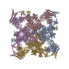
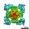
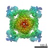
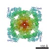
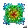
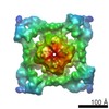
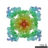
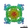
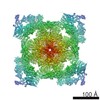
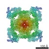
- PDB-5j8v: Structure of rabbit ryanodine receptor RyR1 open state activated ... -
+ Open data

- Basic information
| Entry | Database: PDB / ID: 5j8v | |||||||||||||||
|---|---|---|---|---|---|---|---|---|---|---|---|---|---|---|---|---|
| Title | Structure of rabbit ryanodine receptor RyR1 open state activated by calcium ion | |||||||||||||||
 Components Components | Ryanodine receptor 1 | |||||||||||||||
 Keywords Keywords | TRANSPORT PROTEIN / endoplasmic reticula / skeletal muscles / intracellular calcium ion channel / activated by calcium ion | |||||||||||||||
| Function / homology |  Function and homology information Function and homology informationATP-gated ion channel activity / terminal cisterna / ryanodine receptor complex / ryanodine-sensitive calcium-release channel activity / release of sequestered calcium ion into cytosol by sarcoplasmic reticulum / ossification involved in bone maturation / skin development / cellular response to caffeine / intracellularly gated calcium channel activity / outflow tract morphogenesis ...ATP-gated ion channel activity / terminal cisterna / ryanodine receptor complex / ryanodine-sensitive calcium-release channel activity / release of sequestered calcium ion into cytosol by sarcoplasmic reticulum / ossification involved in bone maturation / skin development / cellular response to caffeine / intracellularly gated calcium channel activity / outflow tract morphogenesis / organelle membrane / toxic substance binding / voltage-gated calcium channel activity / skeletal muscle fiber development / release of sequestered calcium ion into cytosol / sarcoplasmic reticulum membrane / cellular response to calcium ion / sarcoplasmic reticulum / muscle contraction / calcium ion transmembrane transport / calcium channel activity / intracellular calcium ion homeostasis / disordered domain specific binding / protein homotetramerization / transmembrane transporter binding / calmodulin binding / calcium ion binding / ATP binding / membrane / identical protein bindingSimilarity search - Function | |||||||||||||||
| Biological species |  Oryctolagus cuniculus (rabbit) Oryctolagus cuniculus (rabbit) | |||||||||||||||
| Method | ELECTRON MICROSCOPY / single particle reconstruction / cryo EM / Resolution: 4.9 Å | |||||||||||||||
 Authors Authors | Wang, X. / Wei, R. / Yin, C. / Sun, F. | |||||||||||||||
| Funding support |  China, 4items China, 4items
| |||||||||||||||
 Citation Citation | Journal: Cell Res / Year: 2016 Title: Structural insights into Ca(2+)-activated long-range allosteric channel gating of RyR1. Authors: Risheng Wei / Xue Wang / Yan Zhang / Saptarshi Mukherjee / Lei Zhang / Qiang Chen / Xinrui Huang / Shan Jing / Congcong Liu / Shuang Li / Guangyu Wang / Yaofang Xu / Sujie Zhu / Alan J ...Authors: Risheng Wei / Xue Wang / Yan Zhang / Saptarshi Mukherjee / Lei Zhang / Qiang Chen / Xinrui Huang / Shan Jing / Congcong Liu / Shuang Li / Guangyu Wang / Yaofang Xu / Sujie Zhu / Alan J Williams / Fei Sun / Chang-Cheng Yin /  Abstract: Ryanodine receptors (RyRs) are a class of giant ion channels with molecular mass over 2.2 mega-Daltons. These channels mediate calcium signaling in a variety of cells. Since more than 80% of the RyR ...Ryanodine receptors (RyRs) are a class of giant ion channels with molecular mass over 2.2 mega-Daltons. These channels mediate calcium signaling in a variety of cells. Since more than 80% of the RyR protein is folded into the cytoplasmic assembly and the remaining residues form the transmembrane domain, it has been hypothesized that the activation and regulation of RyR channels occur through an as yet uncharacterized long-range allosteric mechanism. Here we report the characterization of a Ca(2+)-activated open-state RyR1 structure by cryo-electron microscopy. The structure has an overall resolution of 4.9 Å and a resolution of 4.2 Å for the core region. In comparison with the previously determined apo/closed-state structure, we observed long-range allosteric gating of the channel upon Ca(2+) activation. In-depth structural analyses elucidated a novel channel-gating mechanism and a novel ion selectivity mechanism of RyR1. Our work not only provides structural insights into the molecular mechanisms of channel gating and regulation of RyRs, but also sheds light on structural basis for channel-gating and ion selectivity mechanisms for the six-transmembrane-helix cation channel family. | |||||||||||||||
| History |
|
- Structure visualization
| Movie |
Movie viewer |
|---|---|
| Structure viewer | Molecule: MolmilJmol/JSmol |
- Downloads & links
-Download
| PDBx/mmCIF format | 5j8v.cif.gz | 2 MB | Display | PDBx/mmCIF format |
|---|---|---|---|---|
| PDB format | pdb5j8v.ent.gz | 1.4 MB | Display | PDB format |
| PDBx/mmJSON format | 5j8v.json.gz | Tree view | PDBx/mmJSON format | |
| Others |  Other downloads Other downloads |
-Validation report
| Arichive directory | https://data.pdbj.org/pub/pdb/validation_reports/j8/5j8vftp://data.pdbj.org/pub/pdb/validation_reports/j8/5j8v | HTTPS FTP |
|---|
-Related structure data
| Related structure data | 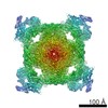 8073MC 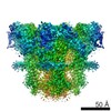 8074C 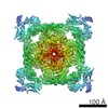 8171C 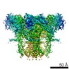 8172C M: map data used to model this data C: citing same article ( |
|---|---|
| Similar structure data |

-Links
 PDBj
PDBj
- Assembly
| Deposited unit | 
|
|---|---|
| 1 |
|
-Components
| #1: Protein | / RyR1 / Skeletal muscle calcium release channel / Skeletal muscle ryanodine receptor / Skeletal ...RyR1 / Skeletal muscle calcium release channel / Skeletal muscle ryanodine receptor / Skeletal muscle-type ryanodine receptor / Type 1 ryanodine receptor Mass: 565908.625 Da / Num. of mol.: 4 / Source method: isolated from a natural source / Source: (natural) Oryctolagus cuniculus (rabbit) / References: UniProt: P11716 |
|---|
-Experimental details
-Experiment
| Experiment | Method: ELECTRON MICROSCOPY |
|---|---|
| EM experiment | Aggregation state: PARTICLE / 3D reconstruction method: single particle reconstruction |
- Sample preparation
| Component | Name: rabbit ryanodine receptor RyR1 / Type: COMPLEX / Entity ID: all / Source: NATURAL | |||||||||||||||||||||||||
|---|---|---|---|---|---|---|---|---|---|---|---|---|---|---|---|---|---|---|---|---|---|---|---|---|---|---|
| Molecular weight | Value: 2.2 MDa / Experimental value: NO | |||||||||||||||||||||||||
| Source (natural) | Organism: Oryctolagus cuniculus (rabbit) | |||||||||||||||||||||||||
| Buffer solution | pH: 7.4 Details: 1:1000 diluted protease inhibitor cocktail was also added in buffer | |||||||||||||||||||||||||
| Buffer component |
| |||||||||||||||||||||||||
| Specimen | Conc.: 5 mg/ml / Embedding applied: NO / Shadowing applied: NO / Staining applied: NO / Vitrification applied: YES | |||||||||||||||||||||||||
| Specimen support | Grid material: COPPER / Grid mesh size: 400 divisions/in. / Grid type: Quantifoil R2/2 | |||||||||||||||||||||||||
| Vitrification | Instrument: FEI VITROBOT MARK IV / Cryogen name: ETHANE / Humidity: 100 % / Chamber temperature: 277 K / Details: Grids were blotted for 2s before plunging. |
- Electron microscopy imaging
| Experimental equipment |  Model: Titan Krios / Image courtesy: FEI Company |
|---|---|
| Microscopy | Model: FEI TITAN KRIOS |
| Electron gun | Electron source: FIELD EMISSION GUN / Accelerating voltage: 300 kV / Illumination mode: FLOOD BEAM |
| Electron lens | Mode: BRIGHT FIELDBright-field microscopy / Nominal magnification: 59000 X / Calibrated magnification: 100286 X / Nominal defocus max: 4000 nm / Nominal defocus min: 1000 nm / Calibrated defocus min: 1300 nm / Calibrated defocus max: 5400 nm / Cs: 2.7 mm / C2 aperture diameter: 50 µm / Alignment procedure: COMA FREE |
| Specimen holder | Cryogen: NITROGEN / Specimen holder model: FEI TITAN KRIOS AUTOGRID HOLDER / Temperature (max): 80 K / Temperature (min): 80 K / Residual tilt: 23 mradians |
| Image recording | Average exposure time: 2 sec. / Electron dose: 48 e/Å2 / Detector mode: INTEGRATING / Film or detector model: FEI FALCON II (4k x 4k) / Num. of grids imaged: 4 / Num. of real images: 4931 |
| Image scans | Sampling size: 14 µm / Width: 4096 / Height: 4096 / Movie frames/image: 31 / Used frames/image: 2-31 |
- Processing
| EM software |
| ||||||||||||||||||||||||||||||||
|---|---|---|---|---|---|---|---|---|---|---|---|---|---|---|---|---|---|---|---|---|---|---|---|---|---|---|---|---|---|---|---|---|---|
| Image processing | Details: The 31 frames of each video were processed into 10 images by merging 3 adjacent frames and then subjected into motion correction using program dosefgpu_driftcorr. | ||||||||||||||||||||||||||||||||
| CTF correction | Type: NONE | ||||||||||||||||||||||||||||||||
| Particle selection | Num. of particles selected: 376537 | ||||||||||||||||||||||||||||||||
| Symmetry | Point symmetry: C4 (4 fold cyclic) | ||||||||||||||||||||||||||||||||
| 3D reconstruction | Resolution: 4.9 Å / Resolution method: FSC 0.143 CUT-OFF / Num. of particles: 41743 / Algorithm: FOURIER SPACE / Num. of class averages: 1 / Symmetry type: POINT | ||||||||||||||||||||||||||||||||
| Atomic model building | Protocol: AB INITIO MODEL / Space: RECIPROCAL | ||||||||||||||||||||||||||||||||
| Atomic model building | PDB-ID: 3J8H Pdb chain-ID: A / Pdb chain residue range: 1-5037 |