 Movie
Movie Controller

[English] 日本語
 Yorodumi
- PDB-5hny: Structural basis of backwards motion in kinesin-14: plus-end dire... -
+ Open data

- Basic information
| Entry | Database: PDB / ID: 5hny | ||||||
|---|---|---|---|---|---|---|---|
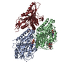
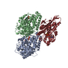
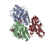
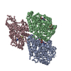
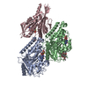
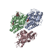
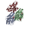
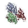
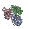
| Title | Structural basis of backwards motion in kinesin-14: plus-end directed nKn669 in the AMPPNP state | ||||||
 Components Components |
| ||||||
 Keywords Keywords |  STRUCTURAL PROTEIN/MOTOR PROTEIN / kinesin / kinesin-14 / microtubule / ATPase / STRUCTURAL PROTEIN-MOTOR PROTEIN complex STRUCTURAL PROTEIN/MOTOR PROTEIN / kinesin / kinesin-14 / microtubule / ATPase / STRUCTURAL PROTEIN-MOTOR PROTEIN complex | ||||||
| Function / homology |  Function and homology information Function and homology informationminus-end directed microtubule sliding / distributive segregation / regulation of mitotic spindle elongation / meiotic spindle assembly / mitotic spindle elongation / mitotic spindle microtubule / meiotic spindle organization / spindle assembly involved in female meiosis / minus-end-directed microtubule motor activity / regulation of mitotic spindle assembly ...minus-end directed microtubule sliding / distributive segregation / regulation of mitotic spindle elongation / meiotic spindle assembly / mitotic spindle elongation / mitotic spindle microtubule / meiotic spindle organization / spindle assembly involved in female meiosis / minus-end-directed microtubule motor activity / regulation of mitotic spindle assembly / microtubule bundle formation / meiotic spindle / mitotic centrosome separation / positive regulation of axon guidance / spindle organization / mitotic spindle assembly / mRNA transport / cytoplasmic microtubule / microtubule-based process / cellular response to interleukin-4 / mitotic spindle organization / chromosome segregation / Hydrolases; Acting on acid anhydrides; Acting on GTP to facilitate cellular and subcellular movement / Hydrolases; Acting on acid anhydrides; Acting on acid anhydrides to facilitate cellular and subcellular movement / structural constituent of cytoskeleton / spindle / microtubule cytoskeleton organization / microtubule cytoskeleton / double-stranded RNA binding / mitotic cell cycle / nervous system development / microtubule binding / microtubule / hydrolase activity / protein heterodimerization activity / cell division / GTPase activity / centrosome / ubiquitin protein ligase binding / GTP binding / protein homodimerization activity / ATP binding / metal ion binding / nucleus / cytosol / cytoplasmSimilarity search - Function | ||||||
| Biological species |  Drosophila melanogaster (fruit fly) Drosophila melanogaster (fruit fly) Rattus norvegicus (Norway rat)Bos taurus (cattle) Rattus norvegicus (Norway rat)Bos taurus (cattle) | ||||||
| Method | ELECTRON MICROSCOPY / helical reconstruction / cryo EM / Resolution: 6.3 Å | ||||||
 Authors Authors | Shigematsu, H. / Yokoyama, T. / Kikkawa, M. / Shirouzu, M. / Nitta, R. | ||||||
 Citation Citation | Journal: Structure / Year: 2016 Title: Structural Basis of Backwards Motion in Kinesin-1-Kinesin-14 Chimera: Implication for Kinesin-14 Motility. Authors: Masahiko Yamagishi / Hideki Shigematsu / Takeshi Yokoyama / Masahide Kikkawa / Mitsuhiro Sugawa / Mari Aoki / Mikako Shirouzu / Junichiro Yajima / Ryo Nitta /  Abstract: Kinesin-14 is a unique minus-end-directed microtubule-based motor. A swinging motion of a class-specific N-terminal neck helix has been proposed to produce minus-end directionality. However, it is ...Kinesin-14 is a unique minus-end-directed microtubule-based motor. A swinging motion of a class-specific N-terminal neck helix has been proposed to produce minus-end directionality. However, it is unclear how swinging of the neck helix is driven by ATP hydrolysis utilizing the highly conserved catalytic core among all kinesins. Here, using a motility assay, we show that in addition to the neck helix, the conserved five residues at the C-terminal region in kinesin-14, namely the neck mimic, are necessary to give kinesin-1 an ability to reverse its directionality toward the minus end of microtubules. Our structural analyses further demonstrate that the C-terminal neck mimic, in cooperation with conformational changes in the catalytic core during ATP binding, forms a kinesin-14 bundle with the N-terminal neck helix to swing toward the minus end of microtubules. Thus, the neck mimic plays a crucial role in coupling the chemical ATPase reaction with the mechanical cycle to produce the minus-end-directed motility of kinesin-14. | ||||||
| History |
|
- Structure visualization
| Movie |
Movie viewer |
|---|---|
| Structure viewer | Molecule: MolmilJmol/JSmol |
- Downloads & links
-Download
| PDBx/mmCIF format | 5hny.cif.gz | 233.6 KB | Display | PDBx/mmCIF format |
|---|---|---|---|---|
| PDB format | pdb5hny.ent.gz | 187.9 KB | Display | PDB format |
| PDBx/mmJSON format | 5hny.json.gz | Tree view | PDBx/mmJSON format | |
| Others |  Other downloads Other downloads |
-Validation report
| Arichive directory | https://data.pdbj.org/pub/pdb/validation_reports/hn/5hnyftp://data.pdbj.org/pub/pdb/validation_reports/hn/5hny | HTTPS FTP |
|---|
-Related structure data
| Related structure data | 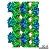 8060MC  8058C  8059C 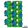 8061C  5hnwC  5hnxC  5hnzC M: map data used to model this data C: citing same article ( |
|---|---|
| Similar structure data |

-Links
 PDBj
PDBj


- Assembly
| Deposited unit | 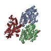
|
|---|---|
| 1 |
|
-Components
-Protein , 3 types, 3 molecules ABK
| #1: Protein | Mass: 48638.793 Da / Num. of mol.: 1 / Fragment: UNP residues 2-439 / Source method: isolated from a natural source / Source: (natural) Bos taurus (cattle) / References: UniProt: P81947 |
|---|---|
| #2: Protein | Mass: 47809.746 Da / Num. of mol.: 1 / Fragment: UNP residues 2-427 / Source method: isolated from a natural source / Source: (natural) Bos taurus (cattle) / References: UniProt: Q6B856 |
| #3: Protein | Mass: 41720.418 Da / Num. of mol.: 1 / Fragment: UNP residues 325-348, UNP residues 664-700 Source method: isolated from a genetically manipulated source Source: (gene. exp.) Drosophila melanogaster (fruit fly), (gene. exp.) Rattus norvegicus (Norway rat)Gene: ncd / Production host:  Escherichia coli (E. coli) / References: UniProt: P20480 Escherichia coli (E. coli) / References: UniProt: P20480 |
-Non-polymers , 5 types, 6 molecules 


| #4: Chemical | ChemComp-GTP / Guanosine triphosphate Mass: 523.180 Da / Num. of mol.: 1 / Source method: obtained synthetically / Formula: C10H16N5O14P3 / Comment: GTP, energy-carrying molecule*YM Mass: 523.180 Da / Num. of mol.: 1 / Source method: obtained synthetically / Formula: C10H16N5O14P3 / Comment: GTP, energy-carrying molecule*YM | ||||||
|---|---|---|---|---|---|---|---|
| #5: Chemical |  Mass: 24.305 Da / Num. of mol.: 2 / Source method: obtained synthetically / Formula: Mg Mass: 24.305 Da / Num. of mol.: 2 / Source method: obtained synthetically / Formula: Mg#6: Chemical | ChemComp-GDP / | Guanosine diphosphate Type: RNA linking / Mass: 443.201 Da / Num. of mol.: 1 / Source method: obtained synthetically / Formula: C10H15N5O11P2 / Comment: GDP, energy-carrying molecule*YM Type: RNA linking / Mass: 443.201 Da / Num. of mol.: 1 / Source method: obtained synthetically / Formula: C10H15N5O11P2 / Comment: GDP, energy-carrying molecule*YM#7: Chemical | ChemComp-TA1 / | Paclitaxel Mass: 853.906 Da / Num. of mol.: 1 / Source method: obtained synthetically / Formula: C47H51NO14 / Comment: medication, chemotherapy*YM Mass: 853.906 Da / Num. of mol.: 1 / Source method: obtained synthetically / Formula: C47H51NO14 / Comment: medication, chemotherapy*YM#8: Chemical | ChemComp-ANP / |  Mass: 506.196 Da / Num. of mol.: 1 / Source method: obtained synthetically / Formula: C10H17N6O12P3 / Comment: AMP-PNP, energy-carrying molecule analogue*YM Mass: 506.196 Da / Num. of mol.: 1 / Source method: obtained synthetically / Formula: C10H17N6O12P3 / Comment: AMP-PNP, energy-carrying molecule analogue*YM |
-Experimental details
-Experiment
| Experiment | Method: ELECTRON MICROSCOPY |
|---|---|
| EM experiment | Aggregation state: FILAMENT / 3D reconstruction method: helical reconstruction |
- Sample preparation
| Component |
| ||||||||||||||||||||||||||||||
|---|---|---|---|---|---|---|---|---|---|---|---|---|---|---|---|---|---|---|---|---|---|---|---|---|---|---|---|---|---|---|---|
| Buffer solution | pH: 6.8 | ||||||||||||||||||||||||||||||
| Specimen | Embedding applied: NO / Shadowing applied: NO / Staining applied: NO / Vitrification applied: YES | ||||||||||||||||||||||||||||||
| Vitrification | Cryogen name: ETHANE |
- Electron microscopy imaging
| Experimental equipment |  Model: Talos Arctica / Image courtesy: FEI Company |
|---|---|
| Microscopy | Model: FEI TECNAI ARCTICA |
| Electron gun | Electron source: FIELD EMISSION GUN / Accelerating voltage: 200 kV / Illumination mode: FLOOD BEAM |
| Electron lens | Mode: BRIGHT FIELDBright-field microscopy |
| Image recording | Electron dose: 30 e/Å2 / Film or detector model: FEI FALCON II (4k x 4k) |
- Processing
| CTF correction | Type: PHASE FLIPPING AND AMPLITUDE CORRECTION |
|---|---|
| Helical symmerty | Angular rotation/subunit: -25.725189 ° / Axial rise/subunit: 8.751208 Å / Axial symmetry: C1 |
| 3D reconstruction | Resolution: 6.3 Å / Resolution method: FSC 0.143 CUT-OFF / Num. of particles: 128254 / Details: High-resolution noise substitution was performed / Symmetry type: HELICAL |
| Atomic model building | Protocol: FLEXIBLE FIT |
