 Movie
Movie Controller

[English] 日本語
 Yorodumi
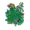
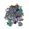
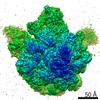
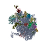
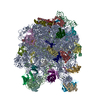
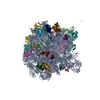
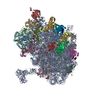
- PDB-4v19: Structure of the large subunit of the mammalian mitoribosome, par... -
+ Open data

- Basic information
| Entry | Database: PDB / ID: 4v19 | |||||||||
|---|---|---|---|---|---|---|---|---|---|---|
| Title | Structure of the large subunit of the mammalian mitoribosome, part 1 of 2 | |||||||||
 Components Components |
| |||||||||
 Keywords Keywords | RIBOSOME / TRANSLATION / LARGE RIBOSOMAL SUBUNIT / MITORIBOSOME / MAMMALIAN MITOCHONDRIAL RIBOSOME / CRYO-EM | |||||||||
| Function / homology |  Function and homology information Function and homology informationMitochondrial translation elongation / Mitochondrial translation termination / rRNA import into mitochondrion / Mitochondrial protein degradation / mitochondrial large ribosomal subunit / organelle membrane / mitochondrial translation / large ribosomal subunit / 5S rRNA binding / cytosolic large ribosomal subunit ...Mitochondrial translation elongation / Mitochondrial translation termination / rRNA import into mitochondrion / Mitochondrial protein degradation / mitochondrial large ribosomal subunit / organelle membrane / mitochondrial translation / large ribosomal subunit / 5S rRNA binding / cytosolic large ribosomal subunit / rRNA binding / ribosome / structural constituent of ribosome / translation / ribonucleoprotein complex / protein domain specific binding / mitochondrion / RNA binding / cytoplasm Similarity search - Function | |||||||||
| Biological species |  | |||||||||
| Method | ELECTRON MICROSCOPY / single particle reconstruction / cryo EM / Resolution: 3.4 Å | |||||||||
 Authors Authors | Greber, B.J. / Boehringer, D. / Leibundgut, M. / Bieri, P. / Leitner, A. / Schmitz, N. / Aebersold, R. / Ban, N. | |||||||||
 Citation Citation | Journal: Nature / Year: 2014 Title: The complete structure of the large subunit of the mammalian mitochondrial ribosome. Authors: Basil J Greber / Daniel Boehringer / Marc Leibundgut / Philipp Bieri / Alexander Leitner / Nikolaus Schmitz / Ruedi Aebersold / Nenad Ban /  Abstract: Mitochondrial ribosomes (mitoribosomes) are extensively modified ribosomes of bacterial descent specialized for the synthesis and insertion of membrane proteins that are critical for energy ...Mitochondrial ribosomes (mitoribosomes) are extensively modified ribosomes of bacterial descent specialized for the synthesis and insertion of membrane proteins that are critical for energy conversion and ATP production inside mitochondria. Mammalian mitoribosomes, which comprise 39S and 28S subunits, have diverged markedly from the bacterial ribosomes from which they are derived, rendering them unique compared to bacterial, eukaryotic cytosolic and fungal mitochondrial ribosomes. We have previously determined at 4.9 Å resolution the architecture of the porcine (Sus scrofa) 39S subunit, which is highly homologous to the human mitoribosomal large subunit. Here we present the complete atomic structure of the porcine 39S large mitoribosomal subunit determined in the context of a stalled translating mitoribosome at 3.4 Å resolution by cryo-electron microscopy and chemical crosslinking/mass spectrometry. The structure reveals the locations and the detailed folds of 50 mitoribosomal proteins, shows the highly conserved mitoribosomal peptidyl transferase active site in complex with its substrate transfer RNAs, and defines the path of the nascent chain in mammalian mitoribosomes along their idiosyncratic exit tunnel. Furthermore, we present evidence that a mitochondrial tRNA has become an integral component of the central protuberance of the 39S subunit where it architecturally substitutes for the absence of the 5S ribosomal RNA, a ubiquitous component of all cytoplasmic ribosomes. | |||||||||
| History |
| |||||||||
| Remark 700 | SHEET DETERMINATION METHOD: DSSP THE SHEETS PRESENTED AS "EA" IN EACH CHAIN ON SHEET RECORDS BELOW ... SHEET DETERMINATION METHOD: DSSP THE SHEETS PRESENTED AS "EA" IN EACH CHAIN ON SHEET RECORDS BELOW IS ACTUALLY AN -6-STRANDED BARREL THIS IS REPRESENTED BY A -5-STRANDED SHEET IN WHICH THE FIRST AND LAST STRANDS ARE IDENTICAL. |
- Structure visualization
| Movie |
Movie viewer |
|---|---|
| Structure viewer | Molecule: MolmilJmol/JSmol |
- Downloads & links
-Download
| PDBx/mmCIF format | 4v19.cif.gz | 1.7 MB | Display | PDBx/mmCIF format |
|---|---|---|---|---|
| PDB format | pdb4v19.ent.gz | 1.4 MB | Display | PDB format |
| PDBx/mmJSON format | 4v19.json.gz | Tree view | PDBx/mmJSON format | |
| Others |  Other downloads Other downloads |
-Validation report
| Summary document | 4v19_validation.pdf.gz | 1.2 MB | Display | wwPDB validaton report |
|---|---|---|---|---|
| Full document | 4v19_full_validation.pdf.gz | 1.3 MB | Display | |
| Data in XML | 4v19_validation.xml.gz | 148.7 KB | Display | |
| Data in CIF | 4v19_validation.cif.gz | 244 KB | Display | |
| Arichive directory | https://data.pdbj.org/pub/pdb/validation_reports/v1/4v19ftp://data.pdbj.org/pub/pdb/validation_reports/v1/4v19 | HTTPS FTP |
-Related structure data
| Related structure data | 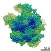 2787MC  4v1aC C: citing same article ( M: map data used to model this data |
|---|---|
| Similar structure data |
-Links
 PDBj
PDBj


- Assembly
| Deposited unit | 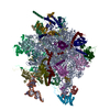
|
|---|---|
| 1 |
|
-Components
+MITORIBOSOMAL ... , 30 types, 30 molecules 0123456789ABDEFIJKNOPQRSTUVWXY
-RNA chain , 1 types, 2 molecules CZ
| #13: RNA chain | Mass: 894.612 Da / Num. of mol.: 2 / Fragment: CCA-3' END / Source method: isolated from a natural source / Details: CHAIN C IS P-SITE TRNA AND CHAIN Z IS A-SITE TRNA / Source: (natural) |
|---|
-Non-polymers , 3 types, 375 molecules 


| #32: Chemical |  Mass: 65.409 Da / Num. of mol.: 2 / Source method: obtained synthetically / Formula: Zn Mass: 65.409 Da / Num. of mol.: 2 / Source method: obtained synthetically / Formula: Zn#33: Chemical | ChemComp-MG /  Mass: 24.305 Da / Num. of mol.: 169 / Source method: obtained synthetically / Formula: Mg Mass: 24.305 Da / Num. of mol.: 169 / Source method: obtained synthetically / Formula: Mg#34: Water | ChemComp-HOH / | Mass: 18.015 Da / Num. of mol.: 204 / Source method: isolated from a natural source / Formula: H2O |
|---|
-Details
| Nonpolymer details | ZINC ION (ZN): ZN IONS COORDINATED BY ZINC FINGER PROTEINS PYRIMIDINE RIBOSIDE-5'-MONOPHOSPHATE ...ZINC ION (ZN): ZN IONS COORDINATE |
|---|---|
| Sequence details | A-SITE TRNA, UNIVERSALLY CONSERVED CCA-3' END P-SITE TRNA, UNIVERSALLY CONSERVED CCA-3' END ...A-SITE TRNA, UNIVERSALL |
-Experimental details
-Experiment
| Experiment | Method: ELECTRON MICROSCOPY |
|---|---|
| EM experiment | Aggregation state: PARTICLE / 3D reconstruction method: single particle reconstruction |
- Sample preparation
| Component | Name: SUS SCROFA 55S MITOCHONDRIAL RIBOSOME / Type: RIBOSOME Details: QUANTIFOIL HOLEY CARBON GRIDS WERE COATED WITH A THIN CARBON FILM |
|---|---|
| Buffer solution | Name: 20 MM HEPES-KOH, 50 MM KCL, 40 MM MGCL2, 1 MM DTT / pH: 7.4 / Details: 20 MM HEPES-KOH, 50 MM KCL, 40 MM MGCL2, 1 MM DTT |
| Specimen | Embedding applied: NO / Shadowing applied: NO / Staining applied: NO / Vitrification applied: YES |
| Specimen support | Details: CARBON |
| Vitrification | Instrument: HOMEMADE PLUNGER / Cryogen name: ETHANE-PROPANE / Details: MIXTURE OF LIQUID ETHANE AND PROPANE |
- Electron microscopy imaging
| Experimental equipment |  Model: Titan Krios / Image courtesy: FEI Company |
|---|---|
| Microscopy | Model: FEI TITAN KRIOS / Date: May 30, 2014 Details: IMAGES WERE ACQUIRED IN 2 SESSIONS ON A FEI TITAN KRIOS IN MAY 2014 |
| Electron gun | Electron source:  FIELD EMISSION GUN / Accelerating voltage: 300 kV / Illumination mode: FLOOD BEAM FIELD EMISSION GUN / Accelerating voltage: 300 kV / Illumination mode: FLOOD BEAM |
| Electron lens | Mode: BRIGHT FIELD / Nominal magnification: 59000 X / Calibrated magnification: 100000 X / Nominal defocus max: 3000 nm / Nominal defocus min: 800 nm / Cs: 2.7 mm |
| Specimen holder | Temperature: 85 K |
| Image recording | Electron dose: 20 e/Å2 / Film or detector model: FEI FALCON II (4k x 4k) |
| Radiation wavelength | Relative weight: 1 |
- Processing
| EM software |
| ||||||||||||||||||||||||||||
|---|---|---|---|---|---|---|---|---|---|---|---|---|---|---|---|---|---|---|---|---|---|---|---|---|---|---|---|---|---|
| CTF correction | Details: PER DETECTOR FRAME | ||||||||||||||||||||||||||||
| Symmetry | Point symmetry: C1 (asymmetric) | ||||||||||||||||||||||||||||
| 3D reconstruction | Method: MAXIMUM LIKELIHOOD BASED REFINEMENT IMPLEMENTED IN RELION Resolution: 3.4 Å / Num. of particles: 141675 / Actual pixel size: 1.4 Å Details: FOR VISUALIZATION PURPOSES THE FINAL MAP WAS FILTERED AND AMPLITUDE CORRECTED IN RELION SUBMISSION BASED ON EXPERIMENTAL DATA FROM EMDB EMD-2787. (DEPOSITION ID: 12830). THE COMBINED ...Details: FOR VISUALIZATION PURPOSES THE FINAL MAP WAS FILTERED AND AMPLITUDE CORRECTED IN RELION SUBMISSION BASED ON EXPERIMENTAL DATA FROM EMDB EMD-2787. (DEPOSITION ID: 12830). THE COMBINED COORDINATES (SPLIT ENTRIES 4V19 AND 4V1A) WERE REFINED IN RECIPROCAL SPACE USING PHENIX.REFINE AGAINST THE MLHL TARGET. FOR THIS, THE CRYO-EM MAPS (EMD-2787) WERE CONVERTED TO RECIPROCAL SPACE STRUCTURE FACTORS. Symmetry type: POINT | ||||||||||||||||||||||||||||
| Atomic model building | Space: REAL | ||||||||||||||||||||||||||||
| Refinement | Highest resolution: 3.4 Å | ||||||||||||||||||||||||||||
| Refinement step | Cycle: LAST / Highest resolution: 3.4 Å
|
