[English] 日本語
Yorodumi
- PDB-4ux1: Cryo-EM structure of antagonist-bound E2P gastric H,K-ATPase (SCH... -

+
Open data


ID or keywords:

Loading...

-
Basic information

Entry
Database: PDB / ID: 4ux1
TitleCryo-EM structure of antagonist-bound E2P gastric H,K-ATPase (SCH.E2. AlF)
Components
  • POTASSIUM-TRANSPORTING ATPASE ALPHA CHAIN 1
  • POTASSIUM-TRANSPORTING ATPASE SUBUNIT BETA
KeywordsTRANSPORT PROTEIN / POTASSIUM-TRANSPORTING ATPASE
Function / homology
Function and homology information


H+/K+-exchanging ATPase / potassium:proton exchanging ATPase complex / P-type potassium:proton transporter activity / Ion transport by P-type ATPases / P-type sodium:potassium-exchanging transporter activity / sodium:potassium-exchanging ATPase complex / sodium ion export across plasma membrane / intracellular potassium ion homeostasis / intracellular sodium ion homeostasis / potassium ion import across plasma membrane ...H+/K+-exchanging ATPase / potassium:proton exchanging ATPase complex / P-type potassium:proton transporter activity / Ion transport by P-type ATPases / P-type sodium:potassium-exchanging transporter activity / sodium:potassium-exchanging ATPase complex / sodium ion export across plasma membrane / intracellular potassium ion homeostasis / intracellular sodium ion homeostasis / potassium ion import across plasma membrane / ATPase activator activity / potassium ion binding / potassium ion transmembrane transport / proton transmembrane transport / cell adhesion / apical plasma membrane / magnesium ion binding / ATP hydrolysis activity / ATP binding / plasma membrane
Similarity search - Function
Gastric H+/K+-transporter P-type ATPase, N-terminal / Gastric H+/K+-ATPase, N terminal domain / Sodium/potassium-transporting ATPase subunit beta / Sodium/potassium-transporting ATPase subunit beta superfamily / Sodium / potassium ATPase beta chain / Sodium and potassium ATPases beta subunits signature 1. / Sodium and potassium ATPases beta subunits signature 2. / P-type ATPase subfamily IIC, subunit alpha / haloacid dehalogenase-like hydrolase / Cation-transporting P-type ATPase, C-terminal ...Gastric H+/K+-transporter P-type ATPase, N-terminal / Gastric H+/K+-ATPase, N terminal domain / Sodium/potassium-transporting ATPase subunit beta / Sodium/potassium-transporting ATPase subunit beta superfamily / Sodium / potassium ATPase beta chain / Sodium and potassium ATPases beta subunits signature 1. / Sodium and potassium ATPases beta subunits signature 2. / P-type ATPase subfamily IIC, subunit alpha / haloacid dehalogenase-like hydrolase / Cation-transporting P-type ATPase, C-terminal / Cation transporting ATPase, C-terminus / Cation transporter/ATPase, N-terminus / Cation-transporting P-type ATPase, N-terminal / Cation transporter/ATPase, N-terminus / Cation transport ATPase (P-type) / E1-E2 ATPase / P-type ATPase, haloacid dehalogenase domain / P-type ATPase, phosphorylation site / P-type ATPase, cytoplasmic domain N / E1-E2 ATPases phosphorylation site. / P-type ATPase, A domain superfamily / P-type ATPase / P-type ATPase, transmembrane domain superfamily / haloacid dehalogenase-like hydrolase / HAD superfamily / HAD-like superfamily
Similarity search - Domain/homology
Potassium-transporting ATPase subunit beta / Potassium-transporting ATPase alpha chain 1
Similarity search - Component
Biological speciesSUS SCROFA (pig)
MethodELECTRON CRYSTALLOGRAPHY / electron crystallography / cryo EM / Resolution: 8 Å
AuthorsAbe, K. / Tani, K. / Fujiyoshi, Y.
Citation
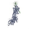
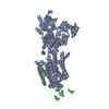
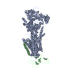
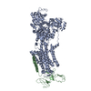
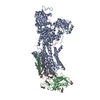
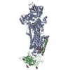
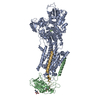
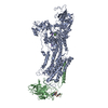
Journal: J Biol Chem / Year: 2014
Title: Systematic comparison of molecular conformations of H+,K+-ATPase reveals an important contribution of the A-M2 linker for the luminal gating.
Authors: Kazuhiro Abe / Kazutoshi Tani / Yoshinori Fujiyoshi /
Abstract: Gastric H(+),K(+)-ATPase, an ATP-driven proton pump responsible for gastric acidification, is a molecular target for anti-ulcer drugs. Here we show its cryo-electron microscopy (EM) structure in an ...Gastric H(+),K(+)-ATPase, an ATP-driven proton pump responsible for gastric acidification, is a molecular target for anti-ulcer drugs. Here we show its cryo-electron microscopy (EM) structure in an E2P analog state, bound to magnesium fluoride (MgF), and its K(+)-competitive antagonist SCH28080, determined at 7 Å resolution by electron crystallography of two-dimensional crystals. Systematic comparison with other E2P-related cryo-EM structures revealed that the molecular conformation in the (SCH)E2·MgF state is remarkably distinguishable. Although the azimuthal position of the A domain of the (SCH)E2·MgF state is similar to that in the E2·AlF (aluminum fluoride) state, in which the transmembrane luminal gate is closed, the arrangement of transmembrane helices in the (SCH)E2·MgF state shows a luminal-open conformation imposed on by bound SCH28080 at its luminal cavity, based on observations of the structure in the SCH28080-bound E2·BeF (beryllium fluoride) state. The molecular conformation of the (SCH)E2·MgF state thus represents a mixed overall structure in which its cytoplasmic and luminal half appear to be independently modulated by a phosphate analog and an antagonist bound to the respective parts of the enzyme. Comparison of the molecular conformations revealed that the linker region connecting the A domain and the transmembrane helix 2 (A-M2 linker) mediates the regulation of luminal gating. The mechanistic rationale underlying luminal gating observed in H(+),K(+)-ATPase is consistent with that observed in sarcoplasmic reticulum Ca(2+)-ATPase and other P-type ATPases and is most likely conserved for the P-type ATPase family in general.
History
DepositionAug 18, 2014Deposition site: PDBE / Processing site: PDBE
Revision 1.0Sep 17, 2014Provider: repository / Type: Initial release
Revision 1.1Sep 24, 2014Group: Database references
Revision 1.2Oct 1, 2014Group: Database references
Revision 1.3Nov 12, 2014Group: Database references

-
Structure visualization

Movie
  • Deposited structure unit
  • Imaged by Jmol
  • Download
  • Simplified surface model + fitted atomic model
  • EMDB-2759
  • Imaged by Jmol
  • Download
  • Superimposition on EM map
  • EMDB-2759
  • Imaged by UCSF Chimera
  • Download
  • Superimposition on EM map
  • EMDB-2759
  • Imaged by UCSF Chimera
  • Download
Movie viewer
Structure viewerMolecule:
MolmilJmol/JSmol

Downloads & links

-
Assembly

Deposited unit
A: POTASSIUM-TRANSPORTING ATPASE ALPHA CHAIN 1
B: POTASSIUM-TRANSPORTING ATPASE SUBUNIT BETA


Theoretical massNumber of molelcules
Total (without water)147,5142
Polymers147,5142
Non-polymers00
Water0
1


  • Idetical with deposited unit
  • defined by software
TypeNameSymmetry operationNumber
identity operation1_555x,y,z1
MethodPISA
Unit cell
Length a, b, c (Å)140.900, 111.300, 320.000
Angle α, β, γ (deg.)90.00, 90.00, 90.00
Int Tables number19
Space group name H-MP212121

-
Components

#1: Protein POTASSIUM-TRANSPORTING ATPASE ALPHA CHAIN 1 / GASTRIC H(+)/K(+) ATPASE SUBUNIT ALPHA / PROTON PUMP / H / K-ATPASE


Mass: 114399.688 Da / Num. of mol.: 1 / Source method: isolated from a natural source / Source: (natural) SUS SCROFA (pig) / References: UniProt: P19156, H+/K+-exchanging ATPase
#2: Protein POTASSIUM-TRANSPORTING ATPASE SUBUNIT BETA / GASTRIC H(+)/K(+) ATPASE SUBUNIT BETA / PROTON PUMP BETA CHA IN / GP60-90 / H / K-ATPASE


Mass: 33113.844 Da / Num. of mol.: 1 / Source method: isolated from a natural source / Source: (natural) SUS SCROFA (pig) / References: UniProt: P18434

-
Experimental details

-
Experiment

ExperimentMethod: ELECTRON CRYSTALLOGRAPHY / Number of used crystals: 267
EM experimentAggregation state: 2D ARRAY / 3D reconstruction method: electron crystallography

-
Sample preparation

ComponentName: antagonist-bound E2P gastric H,K-ATPase / Type: COMPLEX
Buffer solutionpH: 4.8
SpecimenEmbedding applied: NO / Shadowing applied: NO / Staining applied: NO / Vitrification applied: YES
VitrificationInstrument: LEICA KF80 / Cryogen name: NITROGEN
Crystal growpH: 4.8 / Details: pH 4.8

-
Data collection

MicroscopyModel: JEOL KYOTO-3000SFF / Date: Oct 29, 2013
Electron gunElectron source: FIELD EMISSION GUN / Accelerating voltage: 300 kV / Illumination mode: FLOOD BEAM
Electron lensMode: BRIGHT FIELDBright-field microscopy / Nominal magnification: 40000 X / Nominal defocus max: 2997 nm / Nominal defocus min: 808 nm
Image recordingElectron dose: 20 e/Å2 / Film or detector model: KODAK SO-163 FILM
DiffractionMean temperature: 4 K
DetectorDate: Oct 29, 2013
Radiation wavelengthRelative weight: 1
ReflectionResolution: 8→129 Å / Num. obs: 35798 / % possible obs: 52.6 % / Biso Wilson estimate: 32.4 Å2

-
Processing

Software
NameVersionClassification
MRCmodel building
CNS1.3refinement
MRCSUITEdata scaling
MRCphasing
3D reconstructionResolution: 8 Å / Resolution method: DIFFRACTION PATTERN/LAYERLINES / Symmetry type: 2D CRYSTAL
RefinementResolution: 8→128.95 Å / Rfactor Rfree error: 0.043 / Data cutoff high absF: 876135.35 / Data cutoff low absF: 0 / Isotropic thermal model: RESTRAINED / Cross valid method: THROUGHOUT / σ(F): 0
Details: BULK SOLVENT MODEL USED SUBMISSION BASED ON EXPERIMENTAL DATA FROM EMDB EMD-2759. (DEPOSITION ID: 12786).
RfactorNum. reflection% reflectionSelection details
Rfree0.516 146 4.9 %RANDOM
Rwork0.507 ---
obs0.507 2994 52.6 %-
Solvent computationSolvent model: FLAT MODEL / Bsol: 4098.69 Å2 / ksol: 0.1 e/Å3
Displacement parametersBiso mean: 0 Å2
Baniso -1Baniso -2Baniso -3
1-25.76 Å20 Å20 Å2
2--37.87 Å20 Å2
3----63.64 Å2
Refine analyze
FreeObs
Luzzati coordinate error1.4 Å2.23 Å
Luzzati d res low-10 Å
Luzzati sigma a0.2 Å-2.74 Å
Refinement stepCycle: LAST / Resolution: 8→128.95 Å
ProteinNucleic acidLigandSolventTotal
Num. atoms9161 0 0 0 9161
Refine LS restraints
Refine-IDTypeDev idealDev ideal target
ELECTRON CRYSTALLOGRAPHYc_bond_d0.024
ELECTRON CRYSTALLOGRAPHYc_bond_d_na
ELECTRON CRYSTALLOGRAPHYc_bond_d_prot
ELECTRON CRYSTALLOGRAPHYc_angle_d
ELECTRON CRYSTALLOGRAPHYc_angle_d_na
ELECTRON CRYSTALLOGRAPHYc_angle_d_prot
ELECTRON CRYSTALLOGRAPHYc_angle_deg2.8
ELECTRON CRYSTALLOGRAPHYc_angle_deg_na
ELECTRON CRYSTALLOGRAPHYc_angle_deg_prot
ELECTRON CRYSTALLOGRAPHYc_dihedral_angle_d25.1
ELECTRON CRYSTALLOGRAPHYc_dihedral_angle_d_na
ELECTRON CRYSTALLOGRAPHYc_dihedral_angle_d_prot
ELECTRON CRYSTALLOGRAPHYc_improper_angle_d5.51
ELECTRON CRYSTALLOGRAPHYc_improper_angle_d_na
ELECTRON CRYSTALLOGRAPHYc_improper_angle_d_prot
ELECTRON CRYSTALLOGRAPHYc_mcbond_it01.5
ELECTRON CRYSTALLOGRAPHYc_mcangle_it02
ELECTRON CRYSTALLOGRAPHYc_scbond_it02
ELECTRON CRYSTALLOGRAPHYc_scangle_it02.5
Refine LS restraints NCSNCS model details: NONE
LS refinement shellResolution: 8→8.5 Å / Rfactor Rfree error: 0.224 / Total num. of bins used: 6
RfactorNum. reflection% reflection
Rfree0.633 8 3 %
Rwork0.51 262 -
obs--29.4 %
Xplor fileSerial no: 1 / Param file: PROTEIN_REP.PARAM / Topol file: PROTEIN.TOP

+
About Yorodumi

-
News

-
Feb 9, 2022. New format data for meta-information of EMDB entries

New format data for meta-information of EMDB entries

  • Version 3 of the EMDB header file is now the official format.
  • The previous official version 1.9 will be removed from the archive.

Related info.:EMDB header

External links:wwPDB to switch to version 3 of the EMDB data model

-
Aug 12, 2020. Covid-19 info

Covid-19 info

URL: https://pdbj.org/emnavi/covid19.php

New page: Covid-19 featured information page in EM Navigator.

Related info.:Covid-19 info / Mar 5, 2020. Novel coronavirus structure data

+
Mar 5, 2020. Novel coronavirus structure data

Novel coronavirus structure data

Related info.:Yorodumi Speices / Aug 12, 2020. Covid-19 info

External links:COVID-19 featured content - PDBj / Molecule of the Month (242):Coronavirus Proteases

+
Jan 31, 2019. EMDB accession codes are about to change! (news from PDBe EMDB page)

EMDB accession codes are about to change! (news from PDBe EMDB page)

  • The allocation of 4 digits for EMDB accession codes will soon come to an end. Whilst these codes will remain in use, new EMDB accession codes will include an additional digit and will expand incrementally as the available range of codes is exhausted. The current 4-digit format prefixed with “EMD-” (i.e. EMD-XXXX) will advance to a 5-digit format (i.e. EMD-XXXXX), and so on. It is currently estimated that the 4-digit codes will be depleted around Spring 2019, at which point the 5-digit format will come into force.
  • The EM Navigator/Yorodumi systems omit the EMD- prefix.

Related info.:Q: What is EMD? / ID/Accession-code notation in Yorodumi/EM Navigator

External links:EMDB Accession Codes are Changing Soon! / Contact to PDBj

+
Jul 12, 2017. Major update of PDB

Major update of PDB

  • wwPDB released updated PDB data conforming to the new PDBx/mmCIF dictionary.
  • This is a major update changing the version number from 4 to 5, and with Remediation, in which all the entries are updated.
  • In this update, many items about electron microscopy experimental information are reorganized (e.g. em_software).
  • Now, EM Navigator and Yorodumi are based on the updated data.

External links:wwPDB Remediation / Enriched Model Files Conforming to OneDep Data Standards Now Available in the PDB FTP Archive

-
Yorodumi

Thousand views of thousand structures

  • Yorodumi is a browser for structure data from EMDB, PDB, SASBDB, etc.
  • This page is also the successor to EM Navigator detail page, and also detail information page/front-end page for Omokage search.
  • The word "yorodu" (or yorozu) is an old Japanese word meaning "ten thousand". "mi" (miru) is to see.

Related info.:EMDB / PDB / SASBDB / Comparison of 3 databanks / Yorodumi Search / Aug 31, 2016. New EM Navigator & Yorodumi / Yorodumi Papers / Jmol/JSmol / Function and homology information / Changes in new EM Navigator and Yorodumi

Read more