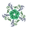
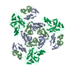
- PDB-4pt2: Myxococcus xanthus encapsulin protein (EncA) -
+
Open data
ID or keywords:
Loading...
-
Basic information
Entry
Database: PDB / ID: 4pt2
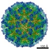
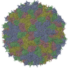
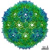
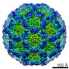
Title
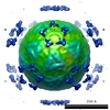
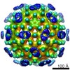
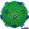
Myxococcus xanthus encapsulin protein (EncA)
Components
Encapsulin protein
Keywords
VIRUS LIKE PARTICLE / HK97 fold / Shell protein
Function / homology
Type 1 encapsulin shell protein / Encapsulating protein for peroxidase / encapsulin nanocompartment / iron ion transport / intracellular iron ion homeostasis / Type 1 encapsulin shell protein EncA
Function and homology information
Biological species
Myxococcus xanthus (bacteria)
Method
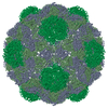
ELECTRON MICROSCOPY / single particle reconstruction / cryo EM / Resolution: 4.6 Å
Journal: EMBO J / Year: 2014 Title: A virus capsid-like nanocompartment that stores iron and protects bacteria from oxidative stress. Authors: Colleen A McHugh / Juan Fontana / Daniel Nemecek / Naiqian Cheng / Anastasia A Aksyuk / J Bernard Heymann / Dennis C Winkler / Alan S Lam / Joseph S Wall / Alasdair C Steven / Egbert Hoiczyk / Abstract: Living cells compartmentalize materials and enzymatic reactions to increase metabolic efficiency. While eukaryotes use membrane-bound organelles, bacteria and archaea rely primarily on protein-bound ...Living cells compartmentalize materials and enzymatic reactions to increase metabolic efficiency. While eukaryotes use membrane-bound organelles, bacteria and archaea rely primarily on protein-bound nanocompartments. Encapsulins constitute a class of nanocompartments widespread in bacteria and archaea whose functions have hitherto been unclear. Here, we characterize the encapsulin nanocompartment from Myxococcus xanthus, which consists of a shell protein (EncA, 32.5 kDa) and three internal proteins (EncB, 17 kDa; EncC, 13 kDa; EncD, 11 kDa). Using cryo-electron microscopy, we determined that EncA self-assembles into an icosahedral shell 32 nm in diameter (26 nm internal diameter), built from 180 subunits with the fold first observed in bacteriophage HK97 capsid. The internal proteins, of which EncB and EncC have ferritin-like domains, attach to its inner surface. Native nanocompartments have dense iron-rich cores. Functionally, they resemble ferritins, cage-like iron storage proteins, but with a massively greater capacity (~30,000 iron atoms versus ~3,000 in ferritin). Physiological data reveal that few nanocompartments are assembled during vegetative growth, but they increase fivefold upon starvation, protecting cells from oxidative stress through iron sequestration.
History
Deposition
Mar 10, 2014
Deposition site: RCSB / Processing site: RCSB
Revision 1.0
Jul 30, 2014
Provider: repository / Type: Initial release
Revision 1.1
Sep 10, 2014
Group: Database references
Revision 1.2
Jul 18, 2018
Group: Data collection / Category: em_software / Item: _em_software.image_processing_id
Method: reference based / Resolution: 4.6 Å / Resolution method: FSC 0.143 CUT-OFF / Num. of particles: 14000 / Nominal pixel size: 1.102 Å / Actual pixel size: 1.102 Å Details: Final map was calculated dividing particles into two independent data sets Symmetry type: POINT
Refinement
Resolution: 4.6→771.4 Å / SU ML: 1.04 / σ(F): 0 / Phase error: 43.52 / Stereochemistry target values: MLHL Details: Homology model of PDB entry 3DKT (I-TASSER), flexible fitting using MDFF and manual re-building using COOT, refined using structure factors derived from EMD-5917
Rfactor
Num. reflection
% reflection
Rwork
0.3489
-
-
obs
0.3489
9859810
99.83 %
all
-
9859810
-
Solvent computation
Shrinkage radii: 0.9 Å / VDW probe radii: 1.11 Å / Solvent model: FLAT BULK SOLVENT MODEL
Refinement step
Cycle: LAST / Resolution: 4.6→771.4 Å
Protein
Nucleic acid
Ligand
Solvent
Total
Num. atoms
6462
0
0
0
6462
Refine LS restraints
Refine-ID
Type
Dev ideal
Number
ELECTRONMICROSCOPY
f_bond_d
0.006
395640
ELECTRONMICROSCOPY
f_angle_d
1.758
536760
ELECTRONMICROSCOPY
f_dihedral_angle_d
18.807
145800
ELECTRONMICROSCOPY
f_chiral_restr
0.063
59760
ELECTRONMICROSCOPY
f_plane_restr
0.006
70560
LS refinement shell
Resolution (Å)
Rfactor Rfree
Num. reflection Rfree
Rfactor Rwork
Num. reflection Rwork
Refine-ID
% reflection obs (%)
4.6-4.7151
0.5016
143
0.5055
705705
ELECTRONMICROSCOPY
100
4.7151-4.8426
0.4629
143
0.4851
704782
ELECTRONMICROSCOPY
100
4.8426-4.9851
0.4948
143
0.4819
706995
ELECTRONMICROSCOPY
100
4.9851-5.146
0.4369
143
0.4563
703960
ELECTRONMICROSCOPY
100
5.146-5.33
0.4128
142
0.4322
704720
ELECTRONMICROSCOPY
100
5.33-5.5434
0.4453
144
0.4301
705819
ELECTRONMICROSCOPY
100
5.5434-5.7957
0.4411
142
0.4013
705292
ELECTRONMICROSCOPY
100
5.7957-6.1013
0.4242
143
0.4323
705593
ELECTRONMICROSCOPY
100
6.1013-6.4836
0.4141
143
0.392
705092
ELECTRONMICROSCOPY
100
6.4836-6.9843
0.3594
143
0.3629
705117
ELECTRONMICROSCOPY
100
6.9843-7.6872
0.3054
142
0.307
705243
ELECTRONMICROSCOPY
100
7.6872-8.7996
0.3137
143
0.293
706205
ELECTRONMICROSCOPY
100
8.7996-11.0868
0.2532
143
0.2476
701578
ELECTRONMICROSCOPY
100
11.0868-782.4801
0.2506
140
0.2398
691712
ELECTRONMICROSCOPY
98
+
About Yorodumi
-
News
-
Feb 9, 2022. New format data for meta-information of EMDB entries
New format data for meta-information of EMDB entries
Version 3 of the EMDB header file is now the official format.
The previous official version 1.9 will be removed from the archive.
In the structure databanks used in Yorodumi, some data are registered as the other names, "COVID-19 virus" and "2019-nCoV". Here are the details of the virus and the list of structure data.
Jan 31, 2019. EMDB accession codes are about to change! (news from PDBe EMDB page)
EMDB accession codes are about to change! (news from PDBe EMDB page)
The allocation of 4 digits for EMDB accession codes will soon come to an end. Whilst these codes will remain in use, new EMDB accession codes will include an additional digit and will expand incrementally as the available range of codes is exhausted. The current 4-digit format prefixed with “EMD-” (i.e. EMD-XXXX) will advance to a 5-digit format (i.e. EMD-XXXXX), and so on. It is currently estimated that the 4-digit codes will be depleted around Spring 2019, at which point the 5-digit format will come into force.
The EM Navigator/Yorodumi systems omit the EMD- prefix.
Related info.:Q: What is EMD? / ID/Accession-code notation in Yorodumi/EM Navigator
Yorodumi is a browser for structure data from EMDB, PDB, SASBDB, etc.
This page is also the successor to EM Navigator detail page, and also detail information page/front-end page for Omokage search.
The word "yorodu" (or yorozu) is an old Japanese word meaning "ten thousand". "mi" (miru) is to see.
Related info.:EMDB / PDB / SASBDB / Comparison of 3 databanks / Yorodumi Search / Aug 31, 2016. New EM Navigator & Yorodumi / Yorodumi Papers / Jmol/JSmol / Function and homology information / Changes in new EM Navigator and Yorodumi
 Movie
Movie Controller


 Open data
 Basic information
 Components
 Keywords
 Function and homology information
 Authors
 Citation
 Structure visualization
 Downloads & links
 Other downloads
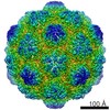


 PDBj
PDBj
 Assembly
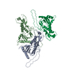

 Sample preparation
 Electron microscopy imaging
 Processing