 Movie
Movie Controller

[English] 日本語
 Yorodumi
- PDB-4ctf: The limits of structural plasticity in a picornavirus capsid reve... -
+ Open data

- Basic information
| Entry | Database: PDB / ID: 4ctf | |||||||||
|---|---|---|---|---|---|---|---|---|---|---|
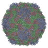
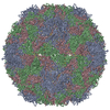
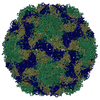
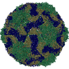
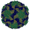
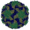
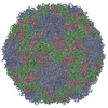
| Title | The limits of structural plasticity in a picornavirus capsid revealed by a massively expanded equine rhinitis A virus particle | |||||||||
 Components Components |
| |||||||||
 Keywords Keywords |  VIRUS / PICORNAVIRUS / CAPSID STRUCTURE / CAPSID EXPANSION / UNCOATING VIRUS / PICORNAVIRUS / CAPSID STRUCTURE / CAPSID EXPANSION / UNCOATING | |||||||||
| Function / homology |  Function and homology information Function and homology informationicosahedral viral capsid / host cell cytoplasm / symbiont entry into host cell / structural molecule activity / virion attachment to host cell / cytoplasmSimilarity search - Function | |||||||||
| Biological species |  EQUINE RHINITIS A VIRUS EQUINE RHINITIS A VIRUS | |||||||||
| Method | ELECTRON MICROSCOPY / single particle reconstruction / cryo EM / Resolution: 17 Å | |||||||||
 Authors Authors | Bakker, S.E. / Groppelli, E. / Pearson, A.R. / Stockley, P.G. / Rowlands, D.J. / Ranson, N.A. | |||||||||
 Citation Citation | Journal: J Virol / Year: 2014 Title: Limits of structural plasticity in a picornavirus capsid revealed by a massively expanded equine rhinitis A virus particle. Authors: Saskia E Bakker / Elisabetta Groppelli / Arwen R Pearson / Peter G Stockley / David J Rowlands / Neil A Ranson /  Abstract: The Picornaviridae family of small, nonenveloped viruses includes major pathogens of humans and animals. They have positive-sense, single-stranded RNA genomes, and the mechanism(s) by which these ...The Picornaviridae family of small, nonenveloped viruses includes major pathogens of humans and animals. They have positive-sense, single-stranded RNA genomes, and the mechanism(s) by which these genomes are introduced into cells to initiate infection remains poorly understood. The structures of presumed uncoating intermediate particles of several picornaviruses show limited expansion and some increased porosity compared to the mature virions. Here, we present the cryo-electron microscopy structure of native equine rhinitis A virus (ERAV), together with the structure of a massively expanded ERAV particle, each at ∼17-Å resolution. The expanded structure has large pores on the particle 3-fold axes and has lost the RNA genome and the capsid protein VP4. The expanded structure thus illustrates both the limits of structural plasticity in such capsids and a plausible route by which genomic RNA might exit. IMPORTANCE: Picornaviruses are important animal and human pathogens that protect their genomic RNAs within a protective protein capsid. Upon infection, this genomic RNA must be able to leave the ...IMPORTANCE: Picornaviruses are important animal and human pathogens that protect their genomic RNAs within a protective protein capsid. Upon infection, this genomic RNA must be able to leave the capsid to initiate a new round of infection. We describe here the structure of a unique, massively expanded state of equine rhinitis A virus that provides insight into how this exit might occur. | |||||||||
| History |
|
- Structure visualization
| Movie |
Movie viewer |
|---|---|
| Structure viewer | Molecule: MolmilJmol/JSmol |
- Downloads & links
-Download
| PDBx/mmCIF format | 4ctf.cif.gz | 7.1 MB | Display | PDBx/mmCIF format |
|---|---|---|---|---|
| PDB format | pdb4ctf.ent.gz | Display | PDB format | |
| PDBx/mmJSON format | 4ctf.json.gz | Tree view | PDBx/mmJSON format | |
| Others |  Other downloads Other downloads |
-Validation report
| Arichive directory | https://data.pdbj.org/pub/pdb/validation_reports/ct/4ctfftp://data.pdbj.org/pub/pdb/validation_reports/ct/4ctf | HTTPS FTP |
|---|
-Related structure data
| Related structure data | 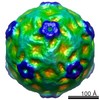 2389MC 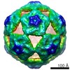 2390C 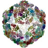 4ctgC M: map data used to model this data C: citing same article ( |
|---|---|
| Similar structure data |

-Links
 PDBj
PDBj
- Assembly
| Deposited unit | 
|
|---|---|
| 1 |
|
-Components
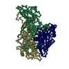
| #1: Protein | Mass: 27296.846 Da / Num. of mol.: 60 / Source method: isolated from a natural source / Details: OHIO HELA CELLS / Source: (natural) EQUINE RHINITIS A VIRUS / References: UniProt: A2TJ51#2: Protein | Mass: 25297.660 Da / Num. of mol.: 60 / Fragment: RESIDUES 81-310 / Source method: isolated from a natural source / Source: (natural) EQUINE RHINITIS A VIRUS / References: UniProt: Q91B42#3: Protein | Mass: 24338.451 Da / Num. of mol.: 60 / Fragment: RESIDUES 311-536 / Source method: isolated from a natural source / Source: (natural) EQUINE RHINITIS A VIRUS / References: UniProt: Q91B37#4: Protein | Mass: 8110.563 Da / Num. of mol.: 60 / Fragment: RESIDUES 1-80 / Source method: isolated from a natural source / Source: (natural) EQUINE RHINITIS A VIRUS / References: UniProt: Q91B42, UniProt: B9VV85*PLUS |
|---|
-Experimental details
-Experiment
| Experiment | Method: ELECTRON MICROSCOPY |
|---|---|
| EM experiment | Aggregation state: PARTICLE / 3D reconstruction method: single particle reconstruction |
- Sample preparation
| Component | Name: EQUINE RHINITIS A VIRUS / Type: VIRUS |
|---|---|
| Specimen | Conc.: 2 mg/ml / Embedding applied: NO / Shadowing applied: NO / Staining applied: NO / Vitrification applied: YES |
| Specimen support | Details: HOLEY CARBON |
| Vitrification | Instrument: HOMEMADE PLUNGER / Cryogen name: ETHANE |
- Electron microscopy imaging
| Experimental equipment |  Model: Tecnai F20 / Image courtesy: FEI Company |
|---|---|
| Microscopy | Model: FEI TECNAI F20 / Date: Jan 1, 2011 |
| Electron gun | Electron source: FIELD EMISSION GUN / Accelerating voltage: 200 kV / Illumination mode: FLOOD BEAM |
| Electron lens | Mode: BRIGHT FIELDBright-field microscopy / Calibrated magnification: 87209 X / Cs: 2 mm |
| Image recording | Electron dose: 15 e/Å2 / Film or detector model: GATAN ULTRASCAN 4000 (4k x 4k) |
| Image scans | Num. digital images: 25 |
| Radiation wavelength | Relative weight: 1 |
- Processing
| EM software |
| ||||||||||||
|---|---|---|---|---|---|---|---|---|---|---|---|---|---|
| CTF correction | Details: PHASE FLIPPING, EACH PARTICLE | ||||||||||||
| Symmetry | Point symmetry: I (icosahedral) | ||||||||||||
| 3D reconstruction | Method: PROJECTION MATCHING / Resolution: 17 Å / Num. of particles: 260 / Nominal pixel size: 1.71 Å / Actual pixel size: 1.71 Å / Symmetry type: POINT | ||||||||||||
| Atomic model building | Protocol: RIGID BODY FIT / Space: REAL / Details: METHOD--RIGID BODY REFINEMENT PROTOCOL--X-RAY | ||||||||||||
| Atomic model building | PDB-ID: 2WFF | ||||||||||||
| Refinement | Highest resolution: 17 Å | ||||||||||||
| Refinement step | Cycle: LAST / Highest resolution: 17 Å
|

