Movie
Movie Controller

[English] 日本語
 Yorodumi
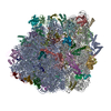
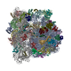
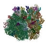
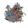
- PDB-3j9y: Cryo-EM structure of tetracycline resistance protein TetM bound t... -
+ Open data

- Basic information
| Entry | Database: PDB / ID: 3j9y | |||||||||
|---|---|---|---|---|---|---|---|---|---|---|
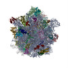
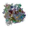
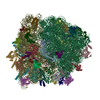
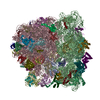
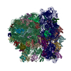
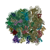
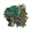
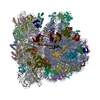
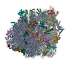
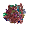
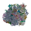
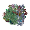
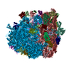
| Title | Cryo-EM structure of tetracycline resistance protein TetM bound to a translating E.coli ribosome | |||||||||
 Components Components |
| |||||||||
 Keywords Keywords | RIBOSOME / antibiotics / protein synthesis / resistance / TetM / tetracycline / tigecycline / translation | |||||||||
| Function / homology |  Function and homology information Function and homology informationribosome disassembly / negative regulation of cytoplasmic translational initiation / stringent response / ornithine decarboxylase inhibitor activity / transcription antitermination factor activity, RNA binding / misfolded RNA binding / Group I intron splicing / RNA folding / transcriptional attenuation / endoribonuclease inhibitor activity ...ribosome disassembly / negative regulation of cytoplasmic translational initiation / stringent response / ornithine decarboxylase inhibitor activity / transcription antitermination factor activity, RNA binding / misfolded RNA binding / Group I intron splicing / RNA folding / transcriptional attenuation / endoribonuclease inhibitor activity / RNA-binding transcription regulator activity / positive regulation of ribosome biogenesis / negative regulation of cytoplasmic translation / four-way junction DNA binding / translational termination / DnaA-L2 complex / translation repressor activity / negative regulation of translational initiation / negative regulation of DNA-templated DNA replication initiation / regulation of mRNA stability / mRNA regulatory element binding translation repressor activity / ribosome assembly / positive regulation of RNA splicing / assembly of large subunit precursor of preribosome / transcription elongation factor complex / cytosolic ribosome assembly / regulation of DNA-templated transcription elongation / DNA endonuclease activity / ribosomal large subunit assembly / transcription antitermination / response to reactive oxygen species / translational initiation / regulation of cell growth / DNA-templated transcription termination / maintenance of translational fidelity / response to radiation / mRNA 5'-UTR binding / large ribosomal subunit / ribosome biogenesis / ribosome binding / regulation of translation / ribosomal small subunit biogenesis / ribosomal small subunit assembly / small ribosomal subunit / small ribosomal subunit rRNA binding / transferase activity / 5S rRNA binding / large ribosomal subunit rRNA binding / cytosolic small ribosomal subunit / cytosolic large ribosomal subunit / cytoplasmic translation / tRNA binding / molecular adaptor activity / rRNA binding / negative regulation of translation / ribosome / structural constituent of ribosome / translation / response to antibiotic / negative regulation of DNA-templated transcription / GTPase activity / mRNA binding / GTP binding / DNA binding / RNA binding / zinc ion binding / membrane / cytoplasm / cytosol Similarity search - Function | |||||||||
| Biological species |   Enterococcus faecalis (bacteria) Enterococcus faecalis (bacteria) | |||||||||
| Method | ELECTRON MICROSCOPY / single particle reconstruction / cryo EM / Resolution: 3.9 Å | |||||||||
 Authors Authors | Arenz, S. / Nguyen, F. / Beckmann, R. / Wilson, D.N. | |||||||||
 Citation Citation | Journal: Proc Natl Acad Sci U S A / Year: 2015 Title: Cryo-EM structure of the tetracycline resistance protein TetM in complex with a translating ribosome at 3.9-Å resolution. Authors: Stefan Arenz / Fabian Nguyen / Roland Beckmann / Daniel N Wilson /  Abstract: Ribosome protection proteins (RPPs) confer resistance to tetracycline by binding to the ribosome and chasing the drug from its binding site. Current models for RPP action are derived from 7.2- to 16- ...Ribosome protection proteins (RPPs) confer resistance to tetracycline by binding to the ribosome and chasing the drug from its binding site. Current models for RPP action are derived from 7.2- to 16-Å resolution structures of RPPs bound to vacant or nontranslating ribosomes. Here we present a cryo-electron microscopy reconstruction of the RPP TetM in complex with a translating ribosome at 3.9-Å resolution. The structure reveals the contacts of TetM with the ribosome, including interaction between the conserved and functionally critical C-terminal extension of TetM with a unique splayed conformation of nucleotides A1492 and A1493 at the decoding center of the small subunit. The resolution enables us to unambiguously model the side chains of the amino acid residues comprising loop III in domain IV of TetM, revealing that the tyrosine residues Y506 and Y507 are not responsible for drug-release as suggested previously but rather for intrafactor contacts that appear to stabilize the conformation of loop III. Instead, Pro509 at the tip of loop III is located directly within the tetracycline binding site where it interacts with nucleotide C1054 of the 16S rRNA, such that RPP action uses Pro509, rather than Y506/Y507, to directly dislodge and release tetracycline from the ribosome. | |||||||||
| History |
|
- Structure visualization
| Movie |
Movie viewer |
|---|---|
| Structure viewer | Molecule: MolmilJmol/JSmol |
- Downloads & links
-Download
| PDBx/mmCIF format | 3j9y.cif.gz | 3.7 MB | Display | PDBx/mmCIF format |
|---|---|---|---|---|
| PDB format | pdb3j9y.ent.gz | Display | PDB format | |
| PDBx/mmJSON format | 3j9y.json.gz | Tree view | PDBx/mmJSON format | |
| Others |  Other downloads Other downloads |
-Validation report
| Summary document | 3j9y_validation.pdf.gz | 1.5 MB | Display | wwPDB validaton report |
|---|---|---|---|---|
| Full document | 3j9y_full_validation.pdf.gz | 1.6 MB | Display | |
| Data in XML | 3j9y_validation.xml.gz | 221.7 KB | Display | |
| Data in CIF | 3j9y_validation.cif.gz | 382.2 KB | Display | |
| Arichive directory | https://data.pdbj.org/pub/pdb/validation_reports/j9/3j9yftp://data.pdbj.org/pub/pdb/validation_reports/j9/3j9y | HTTPS FTP |
-Related structure data
| Related structure data |  6311MC M: map data used to model this data C: citing same article ( |
|---|---|
| Similar structure data |


-Links
 PDBj
PDBj


- Assembly
| Deposited unit | 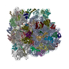
|
|---|---|
| 1 |
|
-Components
-RNA chain , 5 types, 5 molecules avxAB
| #1: RNA chain | Mass: 498909.844 Da / Num. of mol.: 1 / Source method: isolated from a natural source / Source: (natural) |
|---|---|
| #15: RNA chain | Mass: 24978.098 Da / Num. of mol.: 1 / Source method: isolated from a natural source / Source: (natural) |
| #16: RNA chain | Mass: 3492.122 Da / Num. of mol.: 1 / Source method: isolated from a natural source / Source: (natural) |
| #25: RNA chain | Mass: 941521.375 Da / Num. of mol.: 1 / Source method: isolated from a natural source / Source: (natural) |
| #26: RNA chain | Mass: 38813.133 Da / Num. of mol.: 1 / Source method: isolated from a natural source / Source: (natural) |
-30S ribosomal protein ... , 20 types, 20 molecules bdefhklopqrtucgijmns
| #2: Protein | Mass: 26652.557 Da / Num. of mol.: 1 / Source method: isolated from a natural source / Source: (natural) |
|---|---|
| #3: Protein | Mass: 23514.199 Da / Num. of mol.: 1 / Source method: isolated from a natural source / Source: (natural) |
| #4: Protein | Mass: 17629.398 Da / Num. of mol.: 1 / Source method: isolated from a natural source / Source: (natural) |
| #5: Protein | Mass: 15727.512 Da / Num. of mol.: 1 / Source method: isolated from a natural source / Source: (natural) |
| #6: Protein | Mass: 14146.557 Da / Num. of mol.: 1 / Source method: isolated from a natural source / Source: (natural) |
| #7: Protein | Mass: 13870.975 Da / Num. of mol.: 1 / Source method: isolated from a natural source / Source: (natural) |
| #8: Protein | Mass: 13768.157 Da / Num. of mol.: 1 / Source method: isolated from a natural source / Source: (natural) |
| #9: Protein | Mass: 10290.816 Da / Num. of mol.: 1 / Source method: isolated from a natural source / Source: (natural) |
| #10: Protein | Mass: 9207.572 Da / Num. of mol.: 1 / Source method: isolated from a natural source / Source: (natural) |
| #11: Protein | Mass: 9724.491 Da / Num. of mol.: 1 / Source method: isolated from a natural source / Source: (natural) |
| #12: Protein | Mass: 9005.472 Da / Num. of mol.: 1 / Source method: isolated from a natural source / Source: (natural) |
| #13: Protein | Mass: 9708.464 Da / Num. of mol.: 1 / Source method: isolated from a natural source / Source: (natural) |
| #14: Protein | Mass: 8524.039 Da / Num. of mol.: 1 / Source method: isolated from a natural source / Source: (natural) |
| #18: Protein | Mass: 26031.316 Da / Num. of mol.: 1 / Source method: isolated from a natural source / Source: (natural) |
| #19: Protein | Mass: 20055.156 Da / Num. of mol.: 1 / Source method: isolated from a natural source / Source: (natural) |
| #20: Protein | Mass: 14886.270 Da / Num. of mol.: 1 / Source method: isolated from a natural source / Source: (natural) |
| #21: Protein | Mass: 11755.597 Da / Num. of mol.: 1 / Source method: isolated from a natural source / Source: (natural) |
| #22: Protein | Mass: 13128.467 Da / Num. of mol.: 1 / Source method: isolated from a natural source / Source: (natural) |
| #23: Protein | Mass: 11677.637 Da / Num. of mol.: 1 / Source method: isolated from a natural source / Source: (natural) |
| #24: Protein | Mass: 10455.355 Da / Num. of mol.: 1 / Source method: isolated from a natural source / Source: (natural) |
-Protein , 1 types, 1 molecules w
| #17: Protein | Mass: 72542.883 Da / Num. of mol.: 1 Source method: isolated from a genetically manipulated source Source: (gene. exp.) Enterococcus faecalis (bacteria) / Production host: |
|---|
+50S ribosomal protein ... , 32 types, 32 molecules CDEFGHIJKLMNOPQRSTUVWXYZ01234567
-Experimental details
-Experiment
| Experiment | Method: ELECTRON MICROSCOPY |
|---|---|
| EM experiment | Aggregation state: PARTICLE / 3D reconstruction method: single particle reconstruction |
- Sample preparation
| Component |
| ||||||||||||||||
|---|---|---|---|---|---|---|---|---|---|---|---|---|---|---|---|---|---|
| Buffer solution | Name: 50 mM HEPES-KOH, 100 mM KOAc, 25 mM Mg(OAc)2, 6 mM b-mercaptoethanol pH: 7.4 Details: 50 mM HEPES-KOH, 100 mM KOAc, 25 mM Mg(OAc)2, 6 mM b-mercaptoethanol | ||||||||||||||||
| Specimen | Embedding applied: NO / Shadowing applied: NO / Staining applied: NO / Vitrification applied: YES | ||||||||||||||||
| Vitrification | Instrument: FEI VITROBOT MARK IV / Cryogen name: ETHANE / Details: Plunged into liquid ethane (FEI VITROBOT MARK IV) |
- Electron microscopy imaging
| Experimental equipment |  Model: Titan Krios / Image courtesy: FEI Company |
|---|---|
| Microscopy | Model: FEI TITAN KRIOS / Date: Mar 14, 2014 |
| Electron gun | Electron source:  FIELD EMISSION GUN / Accelerating voltage: 300 kV / Illumination mode: SPOT SCAN FIELD EMISSION GUN / Accelerating voltage: 300 kV / Illumination mode: SPOT SCAN |
| Electron lens | Mode: BRIGHT FIELD / Nominal magnification: 125085 X / Nominal defocus max: 3500 nm / Nominal defocus min: 700 nm / Cs: 0 mm |
| Specimen holder | Specimen holder model: FEI TITAN KRIOS AUTOGRID HOLDER |
| Image recording | Electron dose: 28 e/Å2 / Film or detector model: FEI FALCON II (4k x 4k) |
| Image scans | Num. digital images: 2753 |
| Radiation | Protocol: SINGLE WAVELENGTH / Monochromatic (M) / Laue (L): M / Scattering type: x-ray |
| Radiation wavelength | Relative weight: 1 |
- Processing
| EM software |
| ||||||||||||
|---|---|---|---|---|---|---|---|---|---|---|---|---|---|
| CTF correction | Details: Defocus groups | ||||||||||||
| Symmetry | Point symmetry: C1 (asymmetric) | ||||||||||||
| 3D reconstruction | Resolution: 3.9 Å / Resolution method: FSC 0.143 CUT-OFF / Num. of particles: 78186 / Nominal pixel size: 1.108 Å / Actual pixel size: 1.108 Å Details: Since images from microscopy were processed in the absence of spatial frequencies higher than 8 A, an FSC cut-off value of 0.143 was used for average resolution determination of 3.9 A ...Details: Since images from microscopy were processed in the absence of spatial frequencies higher than 8 A, an FSC cut-off value of 0.143 was used for average resolution determination of 3.9 A (Scheres and Chen, 2012). The final map was sharpened by applying an automatically determined negative B-factor using the program EMBFACTOR (Fernandez et al, 2008). (Single particle--Applied symmetry: C1) Symmetry type: POINT | ||||||||||||
| Atomic model building | Protocol: RIGID BODY FIT / Space: REAL / Details: REFINEMENT PROTOCOL--rigid body | ||||||||||||
| Atomic model building | PDB-ID: 5AFI | ||||||||||||
| Refinement step | Cycle: LAST
|