Movie
Movie Controller

[English] 日本語
 Yorodumi
- PDB-3j4p: Electron Microscopy Analysis of a Disaccharide Analog complex Rev... -
+ Open data

- Basic information
| Entry | Database: PDB / ID: 3j4p | |||||||||
|---|---|---|---|---|---|---|---|---|---|---|
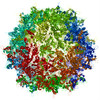
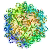
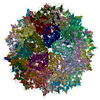
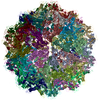
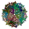
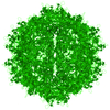
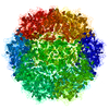
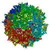
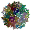
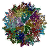
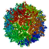
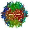
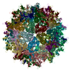
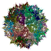
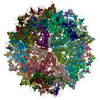
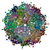
| Title | Electron Microscopy Analysis of a Disaccharide Analog complex Reveals Receptor Interactions of Adeno-Associated Virus | |||||||||
 Components Components | Capsid protein VP1 | |||||||||
 Keywords Keywords | VIRUS / Virus cell-receptor interaction | |||||||||
| Function / homology |  Function and homology information Function and homology informationpermeabilization of host organelle membrane involved in viral entry into host cell / symbiont entry into host cell via permeabilization of inner membrane / host cell nucleolus / T=1 icosahedral viral capsid / clathrin-dependent endocytosis of virus by host cell / structural molecule activity / virion attachment to host cell Similarity search - Function | |||||||||
| Biological species |   Adeno-associated virus Adeno-associated virus | |||||||||
| Method | ELECTRON MICROSCOPY / single particle reconstruction / cryo EM / Resolution: 4.8 Å | |||||||||
 Authors Authors | Xie, Q. / Chapman, M.S. | |||||||||
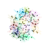
 Citation Citation | Journal: J Struct Biol / Year: 2013 Title: Electron microscopy analysis of a disaccharide analog complex reveals receptor interactions of adeno-associated virus. Authors: Qing Xie / Michael Spilman / Nancy L Meyer / Thomas F Lerch / Scott M Stagg / Michael S Chapman /  Abstract: Mechanistic studies of macromolecular complexes often feature X-ray structures of complexes with bound ligands. The attachment of adeno-associated virus (AAV) to cell surface glycosaminoglycans (GAGs) ...Mechanistic studies of macromolecular complexes often feature X-ray structures of complexes with bound ligands. The attachment of adeno-associated virus (AAV) to cell surface glycosaminoglycans (GAGs) is an example that has not proven amenable to crystallography, because the binding of GAG analogs disrupts lattice contacts. The interactions of AAV with GAGs are of interest in mediating the cell specificity of AAV-based gene therapy vectors. Previous electron microscopy led to differing conclusions on the exact binding site and the existence of large ligand-induced conformational changes in the virus. Conformational changes are expected during cell entry, but it has remained unclear whether the electron microscopy provided evidence of their induction by GAG-binding. Taking advantage of automated data collection, careful processing and new methods of structure refinement, the structure of AAV-DJ complexed with sucrose octasulfate is determined by electron microscopy difference map analysis to 4.8Å resolution. At this higher resolution, individual sulfate groups are discernible, providing a stereochemical validation of map interpretation, and highlighting interactions with two surface arginines that have been implicated in genetic studies. Conformational changes induced by the SOS are modest and limited to the loop most directly interacting with the ligand. While the resolution attainable will depend on sample order and other factors, there are an increasing number of macromolecular complexes that can be studied by cryo-electron microscopy at resolutions beyond 5Å, for which the approaches used here could be used to characterize the binding of inhibitors and other small molecule effectors when crystallography is not tractable. | |||||||||
| History |
|
- Structure visualization
| Movie |
Movie viewer |
|---|---|
| Structure viewer | Molecule: MolmilJmol/JSmol |
- Downloads & links
-Download
| PDBx/mmCIF format | 3j4p.cif.gz | 121 KB | Display | PDBx/mmCIF format |
|---|---|---|---|---|
| PDB format | pdb3j4p.ent.gz | 90.2 KB | Display | PDB format |
| PDBx/mmJSON format | 3j4p.json.gz | Tree view | PDBx/mmJSON format | |
| Others |  Other downloads Other downloads |
-Validation report
| Arichive directory | https://data.pdbj.org/pub/pdb/validation_reports/j4/3j4pftp://data.pdbj.org/pub/pdb/validation_reports/j4/3j4p | HTTPS FTP |
|---|
-Related structure data
| Related structure data | 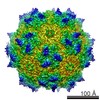 5681MC M: map data used to model this data C: citing same article ( |
|---|---|
| Similar structure data |


-Links
 PDBj
PDBj


- Assembly
| Deposited unit | 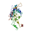
|
|---|---|
| 1 | x 60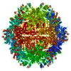
|
| 2 |
|
| 3 | x 5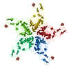
|
| 4 | x 6
|
| 5 | 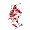
|
| Symmetry | Point symmetry: (Schoenflies symbol: I (icosahedral)) |
-Components
| #1: Protein | Mass: 58577.531 Da / Num. of mol.: 1 / Fragment: SEE REMARK 999 Source method: isolated from a genetically manipulated source Source: (gene. exp.) Adeno-associated virus / Gene: cap / Plasmid: pAVDJ / Production host:   Spodoptera frugiperda (fall armyworm) / Strain (production host): sf9 / References: UniProt: P03135*PLUS Spodoptera frugiperda (fall armyworm) / Strain (production host): sf9 / References: UniProt: P03135*PLUS |
|---|---|
| #2: Polysaccharide | 1,3,4,6-tetra-O-sulfo-beta-D-fructofuranose-(2-1)-2,3,4,6-tetra-O-sulfonato-alpha-D-glucopyranose / sucrose octasulfate  , Oligosaccharide / Class: Substrate analog / Mass: 982.803 Da / Num. of mol.: 1 , Oligosaccharide / Class: Substrate analog / Mass: 982.803 Da / Num. of mol.: 1Source method: isolated from a genetically manipulated source Details: oligosaccharide with reducing-end-to-reducing-end glycosidic bond References: sucrose octasulfate |
| #3: Chemical | ChemComp-NA /   Mass: 22.990 Da / Num. of mol.: 1 / Source method: obtained synthetically / Formula: Na Mass: 22.990 Da / Num. of mol.: 1 / Source method: obtained synthetically / Formula: Na |
| #4: Chemical | ChemComp-MG /   Mass: 24.305 Da / Num. of mol.: 1 / Source method: obtained synthetically / Formula: Mg Mass: 24.305 Da / Num. of mol.: 1 / Source method: obtained synthetically / Formula: Mg |
| #5: Water | ChemComp-HOH / Water Mass: 18.015 Da / Num. of mol.: 1 / Source method: isolated from a natural source / Formula: H2O Mass: 18.015 Da / Num. of mol.: 1 / Source method: isolated from a natural source / Formula: H2O |
| Sequence details | SAMPLE CONTAINED FULL-LENGTH CAPSID PROTEIN, BUT RESIDUES 1-220 WERE NOT MODELED. |
-Experimental details
-Experiment
| Experiment | Method: ELECTRON MICROSCOPY |
|---|---|
| EM experiment | Aggregation state: PARTICLE / 3D reconstruction method: single particle reconstruction |
- Sample preparation
| Component | Name: Recombinant Adeno-Associated Virus DJ / Type: VIRUS |
|---|---|
| Molecular weight | Value: 3.7 MDa / Experimental value: NO |
| Buffer solution | Name: 10 mM Tris, 125 mM NaCl, 1 mM MgCl2 / pH: 6.8 / Details: 10 mM Tris, 125 mM NaCl, 1 mM MgCl2 |
| Specimen | Conc.: 1.5 mg/ml / Embedding applied: NO / Shadowing applied: NO / Staining applied: NO / Vitrification applied: YES |
| Specimen support | Details: 400 mesh carbon-coated grids (C-flat) glow-discharged for 5 seconds (Gatan Model 950) |
| Vitrification | Instrument: FEI VITROBOT MARK IV / Cryogen name: ETHANE / Humidity: 100 % Details: Both sides of the grid were blotted for 2.5 seconds before plunging in liquid ethane (FEI VITROBOT MARK IV). Method: Both sides of the grid were blotted for 2.5 seconds. |
- Electron microscopy imaging
| Experimental equipment |  Model: Titan Krios / Image courtesy: FEI Company |
|---|---|
| Microscopy | Model: FEI TITAN KRIOS / Date: Apr 15, 2013 |
| Electron gun | Electron source: FIELD EMISSION GUN / Accelerating voltage: 120 kV / Illumination mode: FLOOD BEAM |
| Electron lens | Mode: BRIGHT FIELDBright-field microscopy / Nominal magnification: 120000 X / Nominal defocus max: 2000 nm / Nominal defocus min: 800 nm / Cs: 2.7 mm |
| Specimen holder | Specimen holder model: FEI TITAN KRIOS AUTOGRID HOLDER / Specimen holder type: FEI TITAN KRIOS AUTOGRID HOLDER / Temperature: 94 K |
| Image recording | Electron dose: 15 e/Å2 / Film or detector model: GATAN ULTRASCAN 4000 (4k x 4k) |
| Image scans | Num. digital images: 5207 |
| Radiation | Protocol: SINGLE WAVELENGTH / Monochromatic (M) / Laue (L): M / Scattering type: x-ray |
| Radiation wavelength | Relative weight: 1 |
- Processing
| EM software |
| ||||||||||||
|---|---|---|---|---|---|---|---|---|---|---|---|---|---|
| CTF correction | Details: each particle | ||||||||||||
| Symmetry | Point symmetry: I (icosahedral) | ||||||||||||
| 3D reconstruction | Method: Fourier methods / Resolution: 4.8 Å / Resolution method: FSC 0.143 CUT-OFF / Num. of particles: 45000 / Nominal pixel size: 1.3067 Å / Actual pixel size: 1.3067 Å / Details: Final map was amplitude corrected using embfactor. / Symmetry type: POINT | ||||||||||||
| Atomic model building | B value: 25 / Protocol: RIGID BODY FIT / Space: REAL / Target criteria: CC / Details: REFINEMENT PROTOCOL--RIGID BODY | ||||||||||||
| Atomic model building | PDB-ID: 3J1Q Accession code: 3J1Q / Source name: PDB / Type: experimental model | ||||||||||||
| Refinement | Highest resolution: 4.8 Å | ||||||||||||
| Refinement step | Cycle: LAST / Highest resolution: 4.8 Å
|