 Movie
Movie Controller

[English] 日本語
 Yorodumi
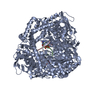
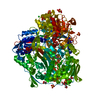
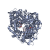
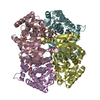
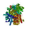
- PDB-1uon: REOVIRUS POLYMERASE LAMBDA-3 LOCALIZED BY ELECTRON CRYOMICROSCOPY... -
+ Open data

- Basic information
| Entry | Database: PDB / ID: 1uon | |||||||||
|---|---|---|---|---|---|---|---|---|---|---|
| Title | REOVIRUS POLYMERASE LAMBDA-3 LOCALIZED BY ELECTRON CRYOMICROSCOPY OF VIRIONS AT 7.6-A RESOLUTION | |||||||||
 Components Components |
| |||||||||
 Keywords Keywords |  POLYMERASE / REOVIRUS / CRYOEM / CORE PROTEIN POLYMERASE / REOVIRUS / CRYOEM / CORE PROTEIN | |||||||||
| Function / homology |  Function and homology information Function and homology informationviral genome replication / viral nucleocapsid / hydrolase activity / RNA-directed RNA polymerase / RNA-dependent RNA polymerase activity / nucleotide binding / RNA bindingSimilarity search - Function | |||||||||
| Biological species |  REOVIRUS REOVIRUSsynthetic construct (others) | |||||||||
| Method | ELECTRON MICROSCOPY / single particle reconstruction / cryo EM / Resolution: 7.6 Å | |||||||||
 Authors Authors | Zhang, X. / Walker, S.B. / Chipman, P.R. / Nibert, M.L. / Baker, T.S. | |||||||||
 Citation Citation | Journal: Nat Struct Biol / Year: 2003 Title: Reovirus polymerase lambda 3 localized by cryo-electron microscopy of virions at a resolution of 7.6 A. Authors: Xing Zhang / Stephen B Walker / Paul R Chipman / Max L Nibert / Timothy S Baker /  Abstract: Reovirus is an icosahedral, double-stranded (ds) RNA virus that uses viral polymerases packaged within the viral core to transcribe its ten distinct plus-strand RNAs. To localize these polymerases, ...Reovirus is an icosahedral, double-stranded (ds) RNA virus that uses viral polymerases packaged within the viral core to transcribe its ten distinct plus-strand RNAs. To localize these polymerases, the structure of the reovirion was refined to a resolution of 7.6 A by cryo-electron microscopy (cryo-EM) and three-dimensional (3D) image reconstruction. X-ray crystal models of reovirus proteins, including polymerase lambda 3, were then fitted into the density map. Each copy of lambda 3 was found anchored to the inner surface of the icosahedral core shell, making major contacts with three molecules of shell protein lambda 1 and overlapping, but not centering on, a five-fold axis. The overlap explains why only one copy of lambda 3 is bound per vertex. lambda 3 is furthermore oriented with its transcript exit channel facing a small channel through the lambda 1 shell, suggesting how the nascent RNA is passed into the large external cavity of the pentameric capping enzyme complex formed by protein lambda 2. | |||||||||
| History |
|
- Structure visualization
| Movie |
Movie viewer |
|---|---|
| Structure viewer | Molecule: MolmilJmol/JSmol |
- Downloads & links
-Download
| PDBx/mmCIF format | 1uon.cif.gz | 272.5 KB | Display | PDBx/mmCIF format |
|---|---|---|---|---|
| PDB format | pdb1uon.ent.gz | 222.2 KB | Display | PDB format |
| PDBx/mmJSON format | 1uon.json.gz | Tree view | PDBx/mmJSON format | |
| Others |  Other downloads Other downloads |
-Validation report
| Arichive directory | https://data.pdbj.org/pub/pdb/validation_reports/uo/1uonftp://data.pdbj.org/pub/pdb/validation_reports/uo/1uon | HTTPS FTP |
|---|
-Related structure data
| Related structure data | |
|---|---|
| Similar structure data |


-Links
 PDBj
PDBj- Assembly
| Deposited unit | 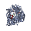
|
|---|---|
| 1 | 
|
-Components
-Protein , 1 types, 1 molecules A
| #1: Protein | Mass: 142423.797 Da / Num. of mol.: 1 / Source method: isolated from a natural source / Source: (natural) REOVIRUS / Strain: T3D / References: UniProt: P17378, UniProt: Q8V1A3*PLUS |
|---|
-RNA chain , 2 types, 2 molecules BC
| #2: RNA chain | Mass: 1681.072 Da / Num. of mol.: 1 / Source method: obtained synthetically / Source: (synth.) synthetic construct (others) |
|---|---|
| #3: RNA chain | Mass: 2461.529 Da / Num. of mol.: 1 / Source method: obtained synthetically / Source: (synth.) synthetic construct (others) |
-Non-polymers , 3 types, 355 molecules 


| #4: Chemical |  Mass: 467.157 Da / Num. of mol.: 3 / Source method: obtained synthetically / Formula: C9H16N3O13P3 Mass: 467.157 Da / Num. of mol.: 3 / Source method: obtained synthetically / Formula: C9H16N3O13P3#5: Chemical |  Mass: 54.938 Da / Num. of mol.: 2 / Source method: obtained synthetically / Formula: Mn Mass: 54.938 Da / Num. of mol.: 2 / Source method: obtained synthetically / Formula: Mn#6: Water | ChemComp-HOH / | WaterMass: 18.015 Da / Num. of mol.: 350 / Source method: isolated from a natural source / Formula: H2O |
|---|
-Experimental details
-Experiment
| Experiment | Method: ELECTRON MICROSCOPY |
|---|---|
| EM experiment | Aggregation state: PARTICLE / 3D reconstruction method: single particle reconstruction |
- Sample preparation
| Component | Name: REOVIRUS POLYMERASE LAMBDA-3 / Type: COMPLEX |
|---|---|
| Buffer solution | Name: TRIS / pH: 7.5 / Details: TRIS |
| Specimen | Conc.: 2 mg/ml / Embedding applied: NO / Shadowing applied: NO / Staining applied: NO / Vitrification applied: YES |
| Specimen support | Details: HOLEY CARBON |
| Crystal grow | *PLUS Method: cryo electron microscopy / Details: cryo electron microscopy |
- Electron microscopy imaging
| EM imaging | Electron source
| ||||||||||||||||||||||||||||||||||||
|---|---|---|---|---|---|---|---|---|---|---|---|---|---|---|---|---|---|---|---|---|---|---|---|---|---|---|---|---|---|---|---|---|---|---|---|---|---|
| Image recording |
| ||||||||||||||||||||||||||||||||||||
| Image scans | Num. digital images: 54 |
- Processing
| EM software | Name: PFT / Category: 3D reconstruction / Details: PFT2D | ||||||||||||
|---|---|---|---|---|---|---|---|---|---|---|---|---|---|
| CTF correction | Details: INDIVIDUAL PARTICLES, PHASE AND AMPLITUDE | ||||||||||||
| Symmetry | Point symmetry: I (icosahedral) | ||||||||||||
| 3D reconstruction | Method: SPHERICAL HARMONICS / Resolution: 7.6 Å / Num. of particles: 7939 / Nominal pixel size: 2.3 Å / Actual pixel size: 2.21 Å / Magnification calibration: CROSS-CORRELATION Details: STRUCTURE OF REOVIRUS CORE (REINISCH ET AL. 2000 NATURE 404:960-967) (PDB ENTRY 1EJ6). PROTEIN ATOMS 9986, NUCLEIC ACID ATOMS 281, HETEROGEN ATOMS 86, SOLVENT ATOMS 350. Symmetry type: POINT | ||||||||||||
| Atomic model building | Protocol: RIGID BODY FIT Details: METHOD--SITUS (CHACON ET AL, 2002 J. MOL. BIOL. 317,375-394) WAS USED TO FIT THE X-RAY STRUCTURE OF REOVIRUS POLYMERASE (TAO ET AL. 2002 CELL 111, 733-745) (PDB CODE 1N35) INTO THE EM ...Details: METHOD--SITUS (CHACON ET AL, 2002 J. MOL. BIOL. 317,375-394) WAS USED TO FIT THE X-RAY STRUCTURE OF REOVIRUS POLYMERASE (TAO ET AL. 2002 CELL 111, 733-745) (PDB CODE 1N35) INTO THE EM DENSITY OF THE 7.6-A RESOLUTION MAP OF REOVIRUS VIRIONS. THE COORDINATES OF THE FITTED REOVIRUS POLYMERASE WERE ALIGNED TO THE X-RAY STRUCTURE OF REOVIRUS CORE (REINISCH ET AL. 2000 NATURE 404,960-967) (PDB ENTRY 1EJ6). ALL MATRICES FOR BUILDING AN ICOSAHEDRON CAN BE FOUND IN 1EJ6. | ||||||||||||
| Atomic model building | PDB-ID: 1UON | ||||||||||||
| Refinement | Highest resolution: 7.6 Å | ||||||||||||
| Refinement step | Cycle: LAST / Highest resolution: 7.6 Å
|
