[English] 日本語
Yorodumi
- PDB-1tge: The structure of immature Dengue virus at 12.5 angstrom -

+
Open data


ID or keywords:

Loading...

-
Basic information

Entry
Database: PDB / ID: 1tge
TitleThe structure of immature Dengue virus at 12.5 angstrom
Componentsenvelope glycoprotein
KeywordsVIRUS / flavivirus / dengue immature virus / prM particle / Icosahedral virus
Function / homology
Function and homology information


flavivirin / serine-type peptidase activity / nucleoside-triphosphate phosphatase / double-stranded RNA binding / clathrin-dependent endocytosis of virus by host cell / viral nucleocapsid / RNA helicase activity / host cell endoplasmic reticulum membrane / protein dimerization activity / RNA helicase ...flavivirin / serine-type peptidase activity / nucleoside-triphosphate phosphatase / double-stranded RNA binding / clathrin-dependent endocytosis of virus by host cell / viral nucleocapsid / RNA helicase activity / host cell endoplasmic reticulum membrane / protein dimerization activity / RNA helicase / fusion of virus membrane with host endosome membrane / viral envelope / virion attachment to host cell / virion membrane / ATP hydrolysis activity / proteolysis / extracellular region / ATP binding / membrane
Similarity search - Function
Flavivirus envelope glycoprotein E, stem/anchor domain / Flavivirus NS3 helicase, C-terminal helical domain / Flavivirus non-structural protein NS2A / Flavivirus non-structural protein NS2A / Flavivirus NS3, petidase S7 / Peptidase S7, Flavivirus NS3 serine protease / Flavivirus NS3 protease (NS3pro) domain profile. / Flavivirus non-structural Protein NS1 / Flavivirus non-structural protein NS1 / Flaviviral glycoprotein E, central domain, subdomain 1 ...Flavivirus envelope glycoprotein E, stem/anchor domain / Flavivirus NS3 helicase, C-terminal helical domain / Flavivirus non-structural protein NS2A / Flavivirus non-structural protein NS2A / Flavivirus NS3, petidase S7 / Peptidase S7, Flavivirus NS3 serine protease / Flavivirus NS3 protease (NS3pro) domain profile. / Flavivirus non-structural Protein NS1 / Flavivirus non-structural protein NS1 / Flaviviral glycoprotein E, central domain, subdomain 1 / Flaviviral glycoprotein E, central domain, subdomain 2 / Flavivirus envelope glycoprotein E, Stem/Anchor domain / Flavivirus glycoprotein E, immunoglobulin-like domain / Flavivirus envelope glycoprotein E, Stem/Anchor domain superfamily / Flavivirus glycoprotein, immunoglobulin-like domain / Flavivirus glycoprotein central and dimerisation domain / Flavivirus glycoprotein, central and dimerisation domains / Flavivirus/Alphavirus glycoprotein, immunoglobulin-like domain superfamily / Flavivirus glycoprotein, central and dimerisation domain superfamily / Flaviviral glycoprotein E, dimerisation domain / DEAD box, Flavivirus / Flavivirus DEAD domain / helicase superfamily c-terminal domain / Immunoglobulin E-set / Superfamilies 1 and 2 helicase C-terminal domain profile. / Superfamilies 1 and 2 helicase ATP-binding type-1 domain profile. / DEAD-like helicases superfamily / Helicase, C-terminal / Helicase superfamily 1/2, ATP-binding domain / Peptidase S1, PA clan / P-loop containing nucleoside triphosphate hydrolase
Similarity search - Domain/homology
Biological speciesDengue virus 2 Puerto Rico/PR159-S1/1969
MethodELECTRON MICROSCOPY / single particle reconstruction / cryo EM / Resolution: 12.5 Å
AuthorsZhang, Y. / Zhang, W. / Ogata, S. / Clements, D. / Strauss, J.H. / Baker, T.S. / Kuhn, R.J. / Rossmann, M.G.
Citation
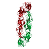
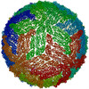
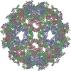
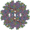
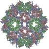
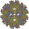
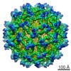
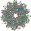
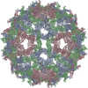
Journal: Structure / Year: 2004
Title: Conformational changes of the flavivirus E glycoprotein.
Authors: Ying Zhang / Wei Zhang / Steven Ogata / David Clements / James H Strauss / Timothy S Baker / Richard J Kuhn / Michael G Rossmann /
Abstract: Dengue virus, a member of the Flaviviridae family, has a surface composed of 180 copies each of the envelope (E) glycoprotein and the membrane (M) protein. The crystal structure of an N-terminal ...Dengue virus, a member of the Flaviviridae family, has a surface composed of 180 copies each of the envelope (E) glycoprotein and the membrane (M) protein. The crystal structure of an N-terminal fragment of E has been determined and compared with a previously described structure. The primary difference between these structures is a 10 degrees rotation about a hinge relating the fusion domain DII to domains DI and DIII. These two rigid body components were used for independent fitting of E into the cryo-electron microscopy maps of both immature and mature dengue viruses. The fitted E structures in these two particles showed a difference of 27 degrees between the two components. Comparison of the E structure in its postfusion state with that in the immature and mature virions shows a rotation approximately around the same hinge. Flexibility of E is apparently a functional requirement for assembly and infection of flaviviruses.
History
DepositionMay 28, 2004Deposition site: RCSB / Processing site: RCSB
Revision 1.0Sep 28, 2004Provider: repository / Type: Initial release
Revision 1.1Apr 30, 2008Group: Version format compliance
Revision 1.2Jul 13, 2011Group: Version format compliance
Revision 1.3Jul 18, 2018Group: Data collection / Category: em_image_scans / em_software / Item: _em_software.image_processing_id / _em_software.name
Revision 1.4Feb 14, 2024Group: Data collection / Database references ...Data collection / Database references / Derived calculations / Refinement description
Category: chem_comp_atom / chem_comp_bond ...chem_comp_atom / chem_comp_bond / database_2 / em_3d_fitting_list / pdbx_initial_refinement_model / pdbx_struct_oper_list / struct_ref_seq_dif
Item: _database_2.pdbx_DOI / _database_2.pdbx_database_accession ..._database_2.pdbx_DOI / _database_2.pdbx_database_accession / _em_3d_fitting_list.accession_code / _em_3d_fitting_list.initial_refinement_model_id / _em_3d_fitting_list.source_name / _em_3d_fitting_list.type / _pdbx_struct_oper_list.name / _pdbx_struct_oper_list.symmetry_operation / _pdbx_struct_oper_list.type / _struct_ref_seq_dif.details
Remark 999SEQUENCE AUTHORS SUBMITTED COORDINATES FOR CA ATOMS ONLY.

-
Structure visualization

Movie
  • Biological unit as complete icosahedral assembly
  • Imaged by Jmol
  • Download
  • Biological unit as icosahedral pentamer
  • Imaged by Jmol
  • Download
  • Biological unit as icosahedral 23 hexamer
  • Imaged by Jmol
  • Download
  • Deposited structure unit
  • Imaged by Jmol
  • Download
  • Superimposition on EM map
  • EMDB-5422
  • Imaged by UCSF Chimera
  • Download
Movie viewer
Structure viewerMolecule:
MolmilJmol/JSmol

Downloads & links

-
Assembly

Deposited unit
A: envelope glycoprotein
B: envelope glycoprotein
C: envelope glycoprotein


Theoretical massNumber of molelcules
Total (without water)131,6293
Polymers131,6293
Non-polymers00
Water0
1
A: envelope glycoprotein
B: envelope glycoprotein
C: envelope glycoprotein
x 60


Theoretical massNumber of molelcules
Total (without water)7,897,759180
Polymers7,897,759180
Non-polymers00
Water0
TypeNameSymmetry operationNumber
identity operation1_555x,y,z1
point symmetry operation59
2


  • Idetical with deposited unit
  • icosahedral asymmetric unit
TypeNameSymmetry operationNumber
identity operation1_555x,y,z1
3
A: envelope glycoprotein
B: envelope glycoprotein
C: envelope glycoprotein
x 5


  • icosahedral pentamer
  • 658 kDa, 15 polymers
Theoretical massNumber of molelcules
Total (without water)658,14715
Polymers658,14715
Non-polymers00
Water0
TypeNameSymmetry operationNumber
identity operation1_555x,y,z1
point symmetry operation4
4
A: envelope glycoprotein
B: envelope glycoprotein
C: envelope glycoprotein
x 6


  • icosahedral 23 hexamer
  • 790 kDa, 18 polymers
Theoretical massNumber of molelcules
Total (without water)789,77618
Polymers789,77618
Non-polymers00
Water0
TypeNameSymmetry operationNumber
identity operation1_555x,y,z1
point symmetry operation5
5


  • Idetical with deposited unit in distinct coordinate
  • icosahedral asymmetric unit, std point frame
TypeNameSymmetry operationNumber
transform to point frame1
SymmetryPoint symmetry: (Hermann–Mauguin notation: 532 / Schoenflies symbol: I (icosahedral))

-
Components

#1: Protein envelope glycoprotein / E glycoprotein / Coordinate model: Cα atoms only


Mass: 43876.441 Da / Num. of mol.: 3 / Source method: isolated from a natural source / Source: (natural) Dengue virus 2 Puerto Rico/PR159-S1/1969 / Genus: Flavivirus / Species: Dengue virus / Strain: PR159/S1 / References: UniProt: P27914

-
Experimental details

-
Experiment

ExperimentMethod: ELECTRON MICROSCOPY
EM experimentAggregation state: PARTICLE / 3D reconstruction method: single particle reconstruction

-
Sample preparation

ComponentName: Dengue-2 immature particle / Type: VIRUS
Details: The samples were produced by adding ammonium chloride to the media in the late infection stage
SpecimenEmbedding applied: NO / Shadowing applied: NO / Staining applied: NO / Vitrification applied: YES

-
Electron microscopy imaging

MicroscopyModel: FEI/PHILIPS CM300FEG/T
Electron gunElectron source: FIELD EMISSION GUN / Illumination mode: FLOOD BEAM
Electron lensMode: BRIGHT FIELDBright-field microscopy / Calibrated magnification: 33000 X / Nominal defocus max: 3640 nm / Nominal defocus min: 1662 nm / Cs: 2 mm
Specimen holderTemperature: 100 K / Tilt angle max: 0 ° / Tilt angle min: 0 °
Image recordingElectron dose: 15 e/Å2 / Film or detector model: KODAK SO-163 FILM

-
Processing

EM software
IDNameCategoryDetails
1EMfitmodel fitting
2POR3D reconstructionJi et al 2003
SymmetryPoint symmetry: I (icosahedral)
3D reconstructionMethod: See primary citation / Resolution: 12.5 Å / Symmetry type: POINT
Atomic model buildingProtocol: OTHER / Space: REAL / Details: METHOD--see primary citation
Atomic model buildingPDB-ID: 1TG8
Accession code: 1TG8 / Source name: PDB / Type: experimental model
Refinement stepCycle: LAST
ProteinNucleic acidLigandSolventTotal
Num. atoms1170 0 0 0 1170

+
About Yorodumi

-
News

-
Feb 9, 2022. New format data for meta-information of EMDB entries

New format data for meta-information of EMDB entries

  • Version 3 of the EMDB header file is now the official format.
  • The previous official version 1.9 will be removed from the archive.

Related info.:EMDB header

External links:wwPDB to switch to version 3 of the EMDB data model

-
Aug 12, 2020. Covid-19 info

Covid-19 info

URL: https://pdbj.org/emnavi/covid19.php

New page: Covid-19 featured information page in EM Navigator.

Related info.:Covid-19 info / Mar 5, 2020. Novel coronavirus structure data

+
Mar 5, 2020. Novel coronavirus structure data

Novel coronavirus structure data

Related info.:Yorodumi Speices / Aug 12, 2020. Covid-19 info

External links:COVID-19 featured content - PDBj / Molecule of the Month (242):Coronavirus Proteases

+
Jan 31, 2019. EMDB accession codes are about to change! (news from PDBe EMDB page)

EMDB accession codes are about to change! (news from PDBe EMDB page)

  • The allocation of 4 digits for EMDB accession codes will soon come to an end. Whilst these codes will remain in use, new EMDB accession codes will include an additional digit and will expand incrementally as the available range of codes is exhausted. The current 4-digit format prefixed with “EMD-” (i.e. EMD-XXXX) will advance to a 5-digit format (i.e. EMD-XXXXX), and so on. It is currently estimated that the 4-digit codes will be depleted around Spring 2019, at which point the 5-digit format will come into force.
  • The EM Navigator/Yorodumi systems omit the EMD- prefix.

Related info.:Q: What is EMD? / ID/Accession-code notation in Yorodumi/EM Navigator

External links:EMDB Accession Codes are Changing Soon! / Contact to PDBj

+
Jul 12, 2017. Major update of PDB

Major update of PDB

  • wwPDB released updated PDB data conforming to the new PDBx/mmCIF dictionary.
  • This is a major update changing the version number from 4 to 5, and with Remediation, in which all the entries are updated.
  • In this update, many items about electron microscopy experimental information are reorganized (e.g. em_software).
  • Now, EM Navigator and Yorodumi are based on the updated data.

External links:wwPDB Remediation / Enriched Model Files Conforming to OneDep Data Standards Now Available in the PDB FTP Archive

-
Yorodumi

Thousand views of thousand structures

  • Yorodumi is a browser for structure data from EMDB, PDB, SASBDB, etc.
  • This page is also the successor to EM Navigator detail page, and also detail information page/front-end page for Omokage search.
  • The word "yorodu" (or yorozu) is an old Japanese word meaning "ten thousand". "mi" (miru) is to see.

Related info.:EMDB / PDB / SASBDB / Comparison of 3 databanks / Yorodumi Search / Aug 31, 2016. New EM Navigator & Yorodumi / Yorodumi Papers / Jmol/JSmol / Function and homology information / Changes in new EM Navigator and Yorodumi

Read more