 Movie
Movie Controller

[English] 日本語
 Yorodumi
- PDB-1mvr: Decoding Center & Peptidyl transferase center from the X-ray stru... -
+ Open data

- Basic information
| Entry | Database: PDB / ID: 1mvr | ||||||
|---|---|---|---|---|---|---|---|
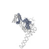
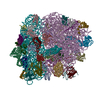
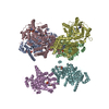
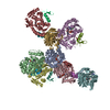
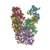
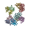
| Title | Decoding Center & Peptidyl transferase center from the X-ray structure of the Thermus thermophilus 70S ribosome, aligned to the low resolution Cryo-EM map of E.coli 70S Ribosome | ||||||
 Components Components |
| ||||||
 Keywords Keywords |  RIBOSOME / RF2 / Release Complex / Conformational Changes RIBOSOME / RF2 / Release Complex / Conformational Changes | ||||||
| Function / homology |  Function and homology information Function and homology informationlarge ribosomal subunit rRNA binding / small ribosomal subunit / cytosolic large ribosomal subunit / tRNA binding / rRNA binding / structural constituent of ribosome / translationSimilarity search - Function | ||||||
| Biological species |  Escherichia coli (E. coli) Escherichia coli (E. coli) | ||||||
| Method | ELECTRON MICROSCOPY / single particle reconstruction / cryo EM / Resolution: 12.8 Å | ||||||
 Authors Authors | Rawat, U.B. / Zavialov, A.V. / Sengupta, J. / Valle, M. / Grassucci, R.A. / Linde, J. / Vestergaard, B. / Ehrenberg, M. / Frank, J. | ||||||
 Citation Citation | Journal: Nature / Year: 2003 Title: A cryo-electron microscopic study of ribosome-bound termination factor RF2. Authors: Urmila B S Rawat / Andrey V Zavialov / Jayati Sengupta / Mikel Valle / Robert A Grassucci / Jamie Linde / Bente Vestergaard / Måns Ehrenberg / Joachim Frank /  Abstract: Protein synthesis takes place on the ribosome, where genetic information carried by messenger RNA is translated into a sequence of amino acids. This process is terminated when a stop codon moves into ...Protein synthesis takes place on the ribosome, where genetic information carried by messenger RNA is translated into a sequence of amino acids. This process is terminated when a stop codon moves into the ribosomal decoding centre (DC) and is recognized by a class-1 release factor (RF). RFs have a conserved GGQ amino-acid motif, which is crucial for peptide release and is believed to interact directly with the peptidyl-transferase centre (PTC) of the 50S ribosomal subunit. Another conserved motif of RFs (SPF in RF2) has been proposed to interact directly with stop codons in the DC of the 30S subunit. The distance between the DC and PTC is approximately 73 A. However, in the X-ray structure of RF2, SPF and GGQ are only 23 A apart, indicating that they cannot be at DC and PTC simultaneously. Here we show that RF2 is in an open conformation when bound to the ribosome, allowing GGQ to reach the PTC while still allowing SPF-stop-codon interaction. The results indicate new interpretations of accuracy in termination, and have implications for how the presence of a stop codon in the DC is signalled to PTC. #1: Journal: Science / Year: 2001Title: Crystal structure of the ribosome at 5.5 A resolution Authors: Yusupov, M.M. / Yusupova, G.Z. / Baucom, A. / Lieberman, K. / Earnest, T.N. / Cate, J.H. / Noller, H. #2: Journal: Cell(Cambridge,Mass.) / Year: 2001Title: The path of messenger RNA through the ribosome Authors: Yusupova, G.Z. / Yusupov, M.M. / Cate, J.H. / Noller, H. | ||||||
| History |
|
- Structure visualization
| Movie |
Movie viewer |
|---|---|
| Structure viewer | Molecule: MolmilJmol/JSmol |
- Downloads & links
-Download
| PDBx/mmCIF format | 1mvr.cif.gz | 30.9 KB | Display | PDBx/mmCIF format |
|---|---|---|---|---|
| PDB format | pdb1mvr.ent.gz | 13.6 KB | Display | PDB format |
| PDBx/mmJSON format | 1mvr.json.gz | Tree view | PDBx/mmJSON format | |
| Others |  Other downloads Other downloads |
-Validation report
| Arichive directory | https://data.pdbj.org/pub/pdb/validation_reports/mv/1mvrftp://data.pdbj.org/pub/pdb/validation_reports/mv/1mvr | HTTPS FTP |
|---|
-Related structure data


-Links
 PDBj
PDBj


- Assembly
| Deposited unit | 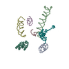
|
|---|---|
| 1 |
|
-Components
-RNA chain , 6 types, 6 molecules 1ABCDE
| #1: RNA chain | Messenger RNA / Coordinate model: P atoms only Mass: 873.540 Da / Num. of mol.: 1 / Source method: isolated from a natural source / Details: based on coordinates from 1GIX, 30S Subunit / Source: (natural) Escherichia coli (E. coli) / Strain: K12 |
|---|---|
| #2: RNA chain | Mass: 14465.605 Da / Num. of mol.: 1 / Source method: isolated from a natural source / Details: based on coordinates from 1GIX, 30S Subunit / Source: (natural) Escherichia coli (E. coli) / Strain: K12 |
| #3: RNA chain | Mass: 31208.639 Da / Num. of mol.: 1 / Source method: isolated from a natural source / Details: based on coordinates from 1GIX, 30S Subunit / Source: (natural) Escherichia coli (E. coli) / Strain: K12 |
| #4: RNA chain | Mass: 6077.673 Da / Num. of mol.: 1 / Source method: isolated from a natural source / Details: based on coordinates from 1GIY, 50S Subunit / Source: (natural) Escherichia coli (E. coli) / Strain: K12 |
| #5: RNA chain | Mass: 18996.311 Da / Num. of mol.: 1 / Source method: isolated from a natural source / Details: based on coordinates from 1GIY, 50S Subunit / Source: (natural) Escherichia coli (E. coli) / Strain: K12 |
| #6: RNA chain | Mass: 8706.199 Da / Num. of mol.: 1 / Source method: isolated from a natural source / Details: based on coordinates from 1GIY, 50S Subunit / Source: (natural) Escherichia coli (E. coli) / Strain: K12 |
-Protein , 2 types, 2 molecules OL
| #7: Protein | / Coordinate model: Cα atoms only Mass: 14920.754 Da / Num. of mol.: 1 / Source method: isolated from a natural source / Details: based on coordinates from 1GIX, 30S Subunit / Source: (natural) Escherichia coli (E. coli) / Strain: K12 / References: UniProt: Q5SHN3 |
|---|---|
| #8: Protein | / Coordinate model: Cα atoms only Mass: 14980.726 Da / Num. of mol.: 1 / Source method: isolated from a natural source / Details: based on coordinates from 1GIY, 50S Subunit / Source: (natural) Escherichia coli (E. coli) / Strain: K12 / References: UniProt: P29395 |
-Experimental details
-Experiment
| Experiment | Method: ELECTRON MICROSCOPY |
|---|---|
| EM experiment | Aggregation state: PARTICLE / 3D reconstruction method: single particle reconstruction |
- Sample preparation
| Component | Name: 70S ribosomeRibosome / Type: RIBOSOME |
|---|---|
| Buffer solution | Name: polymix buffer / pH: 7.5 / Details: polymix buffer |
| Specimen | Conc.: 32 mg/ml / Embedding applied: NO / Shadowing applied: NO / Staining applied: NO / Vitrification applied: YES |
| Vitrification | Instrument: HOMEMADE PLUNGER / Cryogen name: ETHANE / Details: Rapid-freezing in liquid ethane |
- Electron microscopy imaging
| Experimental equipment |  Model: Tecnai F20 / Image courtesy: FEI Company |
|---|---|
| Microscopy | Model: FEI TECNAI F20 / Date: Nov 8, 2001 |
| Electron gun | Electron source: FIELD EMISSION GUN / Accelerating voltage: 200 kV / Illumination mode: FLOOD BEAM |
| Electron lens | Mode: BRIGHT FIELDBright-field microscopy / Nominal magnification: 50000 X / Calibrated magnification: 49696 X / Nominal defocus max: 4500 nm / Nominal defocus min: 2020 nm / Cs: 2 mm |
| Specimen holder | Temperature: 93 K / Tilt angle max: 0 ° / Tilt angle min: 0 ° |
| Image recording | Electron dose: 20 e/Å2 / Film or detector model: KODAK SO-163 FILM |
- Processing
| EM software |
| |||||||||||||||||||||
|---|---|---|---|---|---|---|---|---|---|---|---|---|---|---|---|---|---|---|---|---|---|---|
| CTF correction | Details: Wiener filtering of 3D-maps | |||||||||||||||||||||
| Symmetry | Point symmetry: C1 (asymmetric) | |||||||||||||||||||||
| 3D reconstruction | Method: Reference Bases Alignment / Resolution: 12.8 Å / Num. of particles: 18199 / Actual pixel size: 2.82 Å / Magnification calibration: TMV / Details: SPIDER package / Symmetry type: POINT | |||||||||||||||||||||
| Atomic model building | Protocol: OTHER / Space: REAL / Details: METHOD--Manual | |||||||||||||||||||||
| Atomic model building |
| |||||||||||||||||||||
| Refinement step | Cycle: LAST
|

