 Movie
Movie Controller


+ Open data

- Basic information
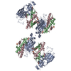
| Entry | Database: PDB / ID: 1fcw | ||||||
|---|---|---|---|---|---|---|---|
| Title | TRNA POSITIONS DURING THE ELONGATION CYCLE | ||||||
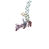
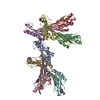
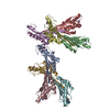
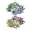
 Components Components | TRNAPHE | ||||||
 Keywords Keywords | RIBOSOME / tRNA binding sites / protein synthesis / elongation cycle | ||||||
| Function / homology | RNA / RNA (> 10) Function and homology information Function and homology information | ||||||
| Biological species |  | ||||||
| Method | ELECTRON MICROSCOPY / single particle reconstruction / cryo EM / Resolution: 17 Å | ||||||
 Authors Authors | Agrawal, R.K. / Spahn, C.M.T. / Penczek, P. / Grassucci, R.A. / Nierhaus, K.H. / Frank, J. | ||||||
 Citation Citation | Journal: J Cell Biol / Year: 2000 Title: Visualization of tRNA movements on the Escherichia coli 70S ribosome during the elongation cycle. Authors: R K Agrawal / C M Spahn / P Penczek / R A Grassucci / K H Nierhaus / J Frank /  Abstract: Three-dimensional cryomaps have been reconstructed for tRNA-ribosome complexes in pre- and posttranslocational states at 17-A resolution. The positions of tRNAs in the A and P sites in the ...Three-dimensional cryomaps have been reconstructed for tRNA-ribosome complexes in pre- and posttranslocational states at 17-A resolution. The positions of tRNAs in the A and P sites in the pretranslocational complexes and in the P and E sites in the posttranslocational complexes have been determined. Of these, the P-site tRNA position is the same as seen earlier in the initiation-like fMet-tRNA(f)(Met)-ribosome complex, where it was visualized with high accuracy. Now, the positions of the A- and E-site tRNAs are determined with similar accuracy. The positions of the CCA end of the tRNAs at the A site are different before and after peptide bond formation. The relative positions of anticodons of P- and E-site tRNAs in the posttranslocational state are such that a codon-anticodon interaction at the E site appears feasible. | ||||||
| History |
|
- Structure visualization
| Movie |
Movie viewer |
|---|---|
| Structure viewer | Molecule: MolmilJmol/JSmol |
- Downloads & links
-Download
| PDBx/mmCIF format | 1fcw.cif.gz | 256.6 KB | Display | PDBx/mmCIF format |
|---|---|---|---|---|
| PDB format | pdb1fcw.ent.gz | 170 KB | Display | PDB format |
| PDBx/mmJSON format | 1fcw.json.gz | Tree view | PDBx/mmJSON format | |
| Others |  Other downloads Other downloads |
-Validation report
| Summary document | 1fcw_validation.pdf.gz | 391 KB | Display | wwPDB validaton report |
|---|---|---|---|---|
| Full document | 1fcw_full_validation.pdf.gz | 741 KB | Display | |
| Data in XML | 1fcw_validation.xml.gz | 72 KB | Display | |
| Data in CIF | 1fcw_validation.cif.gz | 83.8 KB | Display | |
| Arichive directory | https://data.pdbj.org/pub/pdb/validation_reports/fc/1fcwftp://data.pdbj.org/pub/pdb/validation_reports/fc/1fcw | HTTPS FTP |
-Related structure data
| Related structure data | |
|---|---|
| Similar structure data |

-Links
 PDBj
PDBj


- Assembly
| Deposited unit | 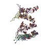
|
|---|---|
| 1 |
|
-Components
| #1: RNA chain | Mass: 24890.121 Da / Num. of mol.: 5 / Source method: isolated from a natural source / Source: (natural) |
|---|
-Experimental details
-Experiment
| Experiment | Method: ELECTRON MICROSCOPY |
|---|---|
| EM experiment | Aggregation state: PARTICLE / 3D reconstruction method: single particle reconstruction |
- Sample preparation
| Component | Name: tRNA-Ribosome complex / Type: COMPLEX / Source: MULTIPLE SOURCES |
|---|---|
| Specimen | Embedding applied: NO / Shadowing applied: NO / Staining applied: NO / Vitrification applied: YES |
| Crystal grow | *PLUS Method: other / Details: electron microscopy |
- Electron microscopy imaging
| Microscopy | Model: FEI/PHILIPS EM420 |
|---|---|
| Electron gun | Electron source: OTHER / Illumination mode: FLOOD BEAM |
| Electron lens | Mode: BRIGHT FIELD / Nominal magnification: 52200 X / Nominal defocus max: 2200 nm / Nominal defocus min: 1200 nm |
| Specimen holder | Specimen holder model: GATAN 626 SINGLE TILT LIQUID NITROGEN CRYO TRANSFER HOLDER |
| Image recording | Electron dose: 10 e/Å2 / Film or detector model: GENERIC FILM |
| Image scans | Scanner model: PERKIN ELMER |
- Processing
| Symmetry | Point symmetry: C1 (asymmetric) | ||||||||||||
|---|---|---|---|---|---|---|---|---|---|---|---|---|---|
| 3D reconstruction | Resolution: 17 Å / Resolution method: FSC 3 SIGMA CUT-OFF / Num. of particles: 15610 / Symmetry type: POINT | ||||||||||||
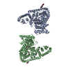
| Atomic model building | Space: REAL Details: DETAILS--TRNA COORDINATES (TRNA09) WERE FIT INTO CRYO-EM MAP AS RIGID BODIES THE RELATIVE POSITIONS OF THE TRNA IS DERIVED BY FITTING THE X-RAY STRUCTURE OF TRNA (TRNA09) INTO THE THREE- ...Details: DETAILS--TRNA COORDINATES (TRNA09) WERE FIT INTO CRYO-EM MAP AS RIGID BODIES THE RELATIVE POSITIONS OF THE TRNA IS DERIVED BY FITTING THE X-RAY STRUCTURE OF TRNA (TRNA09) INTO THE THREE-DIMENSIONAL CRYO-ELECTRON MICROSCOPY MAPS OF FUNCTIONAL COMPLEXES OF THE E. COLI 70S RIBOSOMES. IT SHOULD BE NOTED THAT ALL TRNA-BINDING POSITIONS ARE NOT OCCUPIED SIMULTANEOSLY. THEREFORE, THERE ARE OVERLAPS BETWEEN THE TWO TRNA COORDINATES. IN ORDER TO SEE THE RELATIVE POSITIONS OF VARIOUS RIBOSOMAL DOMAINS, CHAIN C (THE P-SITE TRNA) SHOULD BE ALIGNED WITH OUR EARLIER DEPOSITED P-SITE POSITION (ID CODE 1EGO). | ||||||||||||
| Refinement | Highest resolution: 17 Å | ||||||||||||
| Refinement step | Cycle: LAST / Highest resolution: 17 Å
|
