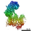
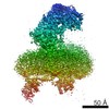
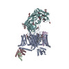
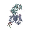
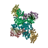
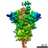
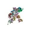
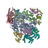
Journal: Nature / Year: 2016 Title: Structure of the voltage-gated calcium channel Ca(v)1.1 at 3.6 Å resolution. Authors: Jianping Wu / Zhen Yan / Zhangqiang Li / Xingyang Qian / Shan Lu / Mengqiu Dong / Qiang Zhou / Nieng Yan / Abstract: The voltage-gated calcium (Ca) channels convert membrane electrical signals to intracellular Ca-mediated events. Among the ten subtypes of Ca channel in mammals, Ca1.1 is specified for the excitation- ...The voltage-gated calcium (Ca) channels convert membrane electrical signals to intracellular Ca-mediated events. Among the ten subtypes of Ca channel in mammals, Ca1.1 is specified for the excitation-contraction coupling of skeletal muscles. Here we present the cryo-electron microscopy structure of the rabbit Ca1.1 complex at a nominal resolution of 3.6 Å. The inner gate of the ion-conducting α1-subunit is closed and all four voltage-sensing domains adopt an 'up' conformation, suggesting a potentially inactivated state. The extended extracellular loops of the pore domain, which are stabilized by multiple disulfide bonds, form a windowed dome above the selectivity filter. One side of the dome provides the docking site for the α2δ-1-subunit, while the other side may attract cations through its negative surface potential. The intracellular I-II and III-IV linker helices interact with the β-subunit and the carboxy-terminal domain of α1, respectively. Classification of the particles yielded two additional reconstructions that reveal pronounced displacement of β and adjacent elements in α1. The atomic model of the Ca1.1 complex establishes a foundation for mechanistic understanding of excitation-contraction coupling and provides a three-dimensional template for molecular interpretations of the functions and disease mechanisms of Ca and Na channels.
History
Deposition
Jul 2, 2016
-
Header (metadata) release
Sep 14, 2016
-
Map release
Sep 14, 2016
-
Update
Jan 1, 2020
-
Current status
Jan 1, 2020
Processing site: PDBj / Status: Released
-
Structure visualization
Movie
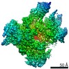
Surface view with section colored by density value
Cryogen name: ETHANE / Chamber humidity: 100 % / Chamber temperature: 281 K / Instrument: FEI VITROBOT MARK IV Details: Grids were blotted for 2 s and flash-frozen in liquid ethane cooled by liquid nitrogen.
Details
This sample was monodisperse.
-
Electron microscopy
Microscope
FEI TITAN KRIOS
Electron beam
Acceleration voltage: 300 kV / Electron source: FIELD EMISSION GUN
Film or detector model: GATAN K2 SUMMIT (4k x 4k) / Detector mode: SUPER-RESOLUTION / Number grids imaged: 5 / Number real images: 9704 / Average exposure time: 8.0 sec. / Average electron dose: 50.0 e/Å2
In the structure databanks used in Yorodumi, some data are registered as the other names, "COVID-19 virus" and "2019-nCoV". Here are the details of the virus and the list of structure data.
Jan 31, 2019. EMDB accession codes are about to change! (news from PDBe EMDB page)
EMDB accession codes are about to change! (news from PDBe EMDB page)
The allocation of 4 digits for EMDB accession codes will soon come to an end. Whilst these codes will remain in use, new EMDB accession codes will include an additional digit and will expand incrementally as the available range of codes is exhausted. The current 4-digit format prefixed with “EMD-” (i.e. EMD-XXXX) will advance to a 5-digit format (i.e. EMD-XXXXX), and so on. It is currently estimated that the 4-digit codes will be depleted around Spring 2019, at which point the 5-digit format will come into force.
The EM Navigator/Yorodumi systems omit the EMD- prefix.
Related info.:Q: What is EMD? / ID/Accession-code notation in Yorodumi/EM Navigator
Yorodumi is a browser for structure data from EMDB, PDB, SASBDB, etc.
This page is also the successor to EM Navigator detail page, and also detail information page/front-end page for Omokage search.
The word "yorodu" (or yorozu) is an old Japanese word meaning "ten thousand". "mi" (miru) is to see.
Related info.:EMDB / PDB / SASBDB / Comparison of 3 databanks / Yorodumi Search / Aug 31, 2016. New EM Navigator & Yorodumi / Yorodumi Papers / Jmol/JSmol / Function and homology information / Changes in new EM Navigator and Yorodumi
 Movie
Movie Controller


 Open data
 Basic information
 Map data
 Sample
 Function and homology information
 L-type voltage-gated calcium channel complex / regulation of calcium ion transmembrane transport via high voltage-gated calcium channel / cellular response to caffeine / calcium ion import across plasma membrane / calcium channel regulator activity /
 Authors
 China, 2 items
  Citation
 Structure visualization
 Downloads & links
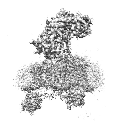 emd_9514.png
 http://ftp.pdbj.org/pub/emdb/structures/EMD-9514
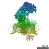


 Sample components
 Processing
 Electron microscopy