National Institutes of Health/National Institute Of Allergy and Infectious Diseases (NIH/NIAID)
R01AI087946
United States
Welch Foundation
Au-1714
United States
National Institutes of Health/National Institute of General Medical Sciences (NIH/NIGMS)
R01GM107629
United States
Citation
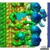
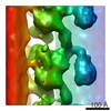
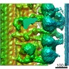
Journal: J Bacteriol / Year: 2017 Title: Imaging the motility and chemotaxis machineries in Helicobacter pylori by cryo-electron tomography. Authors: Zhuan Qin / Wei-Ting Lin / Shiwei Zhu / Aime T Franco / Jun Liu / Abstract: Helicobacter pylori is a bacterial pathogen that can cause many gastrointestinal diseases including ulcers and gastric cancer. A unique chemotaxis-mediated motility is critical for H. pylori to ...Helicobacter pylori is a bacterial pathogen that can cause many gastrointestinal diseases including ulcers and gastric cancer. A unique chemotaxis-mediated motility is critical for H. pylori to colonize in the human stomach and to establish chronic infection, but the underlying molecular mechanisms are not well understood. Here we employ cryo-electron tomography to reveal detailed structures of the H. pylori cell envelope including the sheathed flagella and chemotaxis arrays. Notably, H. pylori possesses a distinctive periplasmic cage-like structure with 18-fold symmetry. We propose that this structure forms a robust platform for recruiting 18 torque generators, which likely provide the higher torque needed for swimming in high-viscosity environments. We also reveal a series of key flagellar assembly intermediates, providing structural evidence that flagellar assembly is tightly coupled with biogenesis of the membrane sheath. Finally, we determine the structure of putative chemotaxis arrays at the flagellar pole, which have implications for how direction of flagellar rotation is regulated. Together, our pilot cryo-ET studies provide novel structural insights into the unipolar flagella of H. pylori and lay a foundation for a better understanding of the unique motility of this organism. IMPORTANCE: Helicobacter pylori is a highly motile bacterial pathogen that colonizes approximately 50% of the world's population. H. pylori can move readily within the viscous mucosal layer of the ...IMPORTANCE: Helicobacter pylori is a highly motile bacterial pathogen that colonizes approximately 50% of the world's population. H. pylori can move readily within the viscous mucosal layer of the stomach. It has become increasingly clear that its unique flagella-driven motility is essential for successful gastric colonization and pathogenesis. Here we use advanced imaging techniques to visualize novel in situ structures with unprecedented detail in intact H. pylori cells. Remarkably, H. pylori possesses multiple unipolar flagella, which are driven by one of the largest flagellar motors found in bacteria. These large motors presumably provide higher torque needed by the bacterial pathogens to navigate in viscous environment of the human stomach.
History
Deposition
Oct 28, 2016
-
Header (metadata) release
Nov 30, 2016
-
Map release
Nov 30, 2016
-
Update
Jan 29, 2020
-
Current status
Jan 29, 2020
Processing site: RCSB / Status: Released
-
Structure visualization
Movie
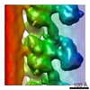
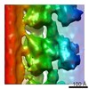
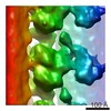
Surface view with section colored by density value
In the structure databanks used in Yorodumi, some data are registered as the other names, "COVID-19 virus" and "2019-nCoV". Here are the details of the virus and the list of structure data.
Jan 31, 2019. EMDB accession codes are about to change! (news from PDBe EMDB page)
EMDB accession codes are about to change! (news from PDBe EMDB page)
The allocation of 4 digits for EMDB accession codes will soon come to an end. Whilst these codes will remain in use, new EMDB accession codes will include an additional digit and will expand incrementally as the available range of codes is exhausted. The current 4-digit format prefixed with “EMD-” (i.e. EMD-XXXX) will advance to a 5-digit format (i.e. EMD-XXXXX), and so on. It is currently estimated that the 4-digit codes will be depleted around Spring 2019, at which point the 5-digit format will come into force.
The EM Navigator/Yorodumi systems omit the EMD- prefix.
Related info.:Q: What is EMD? / ID/Accession-code notation in Yorodumi/EM Navigator
Yorodumi is a browser for structure data from EMDB, PDB, SASBDB, etc.
This page is also the successor to EM Navigator detail page, and also detail information page/front-end page for Omokage search.
The word "yorodu" (or yorozu) is an old Japanese word meaning "ten thousand". "mi" (miru) is to see.
Related info.:EMDB / PDB / SASBDB / Comparison of 3 databanks / Yorodumi Search / Aug 31, 2016. New EM Navigator & Yorodumi / Yorodumi Papers / Jmol/JSmol / Function and homology information / Changes in new EM Navigator and Yorodumi
 Movie
Movie Controller

 Yorodumi
 Open data
 Basic information
 Map data
 Sample

 Helicobacter pylori (bacteria)
 Authors
 United States, 3 items
  Citation
 Structure visualization
 Movie viewer
 Downloads & links
 emd_8460.png
 http://ftp.pdbj.org/pub/emdb/structures/EMD-8460
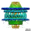
 Sample components
 Processing
 Electron microscopy
