National Institutes of Health/National Institute of General Medical Sciences (NIH/NIGMS)
R01-GM099989
United States
Citation
Journal: J Virol / Year: 2016 Title: Visualization and Sequencing of Membrane Remodeling Leading to Influenza Virus Fusion. Authors: Long Gui / Jamie L Ebner / Alexander Mileant / James A Williams / Kelly K Lee / Abstract: Protein-mediated membrane fusion is an essential step in many fundamental biological events, including enveloped virus infection. The nature of protein and membrane intermediates and the sequence of ...Protein-mediated membrane fusion is an essential step in many fundamental biological events, including enveloped virus infection. The nature of protein and membrane intermediates and the sequence of membrane remodeling during these essential processes remain poorly understood. Here we used cryo-electron tomography (cryo-ET) to image the interplay between influenza virus and vesicles with a range of lipid compositions. By following the population kinetics of membrane fusion intermediates imaged by cryo-ET, we found that membrane remodeling commenced with the hemagglutinin fusion protein spikes grappling onto the target membrane, followed by localized target membrane dimpling as local clusters of hemagglutinin started to undergo conformational refolding. The local dimples then transitioned to extended, tightly apposed contact zones where the two proximal membrane leaflets were in most cases indistinguishable from each other, suggesting significant dehydration and possible intermingling of the lipid head groups. Increasing the content of fusion-enhancing cholesterol or bis-monoacylglycerophosphate in the target membrane led to an increase in extended contact zone formation. Interestingly, hemifused intermediates were found to be extremely rare in the influenza virus fusion system studied here, most likely reflecting the instability of this state and its rapid conversion to postfusion complexes, which increased in population over time. By tracking the populations of fusion complexes over time, the architecture and sequence of membrane reorganization leading to efficient enveloped virus fusion were thus resolved. IMPORTANCE: Enveloped viruses employ specialized surface proteins to mediate fusion of cellular and viral membranes that results in the formation of pores through which the viral genetic material is ...IMPORTANCE: Enveloped viruses employ specialized surface proteins to mediate fusion of cellular and viral membranes that results in the formation of pores through which the viral genetic material is delivered to the cell. For influenza virus, the trimeric hemagglutinin (HA) glycoprotein spike mediates host cell attachment and membrane fusion. While structures of a subset of conformations and parts of the fusion machinery have been characterized, the nature and sequence of membrane deformations during fusion have largely eluded characterization. Building upon studies that focused on early stages of HA-mediated membrane remodeling, here cryo-electron tomography (cryo-ET) was used to image the three-dimensional organization of intact influenza virions at different stages of fusion with liposomes, leading all the way to completion of the fusion reaction. By monitoring the evolution of fusion intermediate populations over the course of acid-induced fusion, we identified the progression of membrane reorganization that leads to efficient fusion by an enveloped virus.
Cryogen name: ETHANE / Chamber humidity: 100 % / Chamber temperature: 25 K / Instrument: FEI VITROBOT MARK IV Details: 3 microliters of solution was added to glow-discharged holey carbon-coated grids (C-flat, 200 mesh, Electron Microscopy Sciences) and plunge-frozen in liquid ethane using an FEI Vitrobot Mark IV..
Sectioning
Other: NO SECTIONING
Fiducial marker
Manufacturer: Electron Microscopy Sciences / Diameter: 10 nm
-
Electron microscopy
Microscope
FEI TECNAI F20
Image recording
Film or detector model: GATAN K2 SUMMIT (4k x 4k) / Detector mode: COUNTING / Digitization - Frames/image: 1-11 / Average exposure time: 1.7 sec. / Average electron dose: 1.5 e/Å2
Electron beam
Acceleration voltage: 200 kV / Electron source: FIELD EMISSION GUN
In the structure databanks used in Yorodumi, some data are registered as the other names, "COVID-19 virus" and "2019-nCoV". Here are the details of the virus and the list of structure data.
Jan 31, 2019. EMDB accession codes are about to change! (news from PDBe EMDB page)
EMDB accession codes are about to change! (news from PDBe EMDB page)
The allocation of 4 digits for EMDB accession codes will soon come to an end. Whilst these codes will remain in use, new EMDB accession codes will include an additional digit and will expand incrementally as the available range of codes is exhausted. The current 4-digit format prefixed with “EMD-” (i.e. EMD-XXXX) will advance to a 5-digit format (i.e. EMD-XXXXX), and so on. It is currently estimated that the 4-digit codes will be depleted around Spring 2019, at which point the 5-digit format will come into force.
The EM Navigator/Yorodumi systems omit the EMD- prefix.
Related info.:Q: What is EMD? / ID/Accession-code notation in Yorodumi/EM Navigator
Yorodumi is a browser for structure data from EMDB, PDB, SASBDB, etc.
This page is also the successor to EM Navigator detail page, and also detail information page/front-end page for Omokage search.
The word "yorodu" (or yorozu) is an old Japanese word meaning "ten thousand". "mi" (miru) is to see.
Related info.:EMDB / PDB / SASBDB / Comparison of 3 databanks / Yorodumi Search / Aug 31, 2016. New EM Navigator & Yorodumi / Yorodumi Papers / Jmol/JSmol / Function and homology information / Changes in new EM Navigator and Yorodumi
 Movie
Movie Controller


 Open data
 Basic information
 Map data
 Sample
 Influenza A virus
 Authors
 United States, 1 items
  Citation
 Structure visualization
 Movie viewer
 Downloads & links
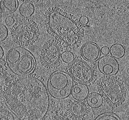 emd_8344.png
 http://ftp.pdbj.org/pub/emdb/structures/EMD-8344
 Sample components
 Processing
 Electron microscopy
 FIELD EMISSION GUN
