Movie
Movie Controller

[English] 日本語
 Yorodumi
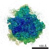
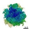
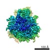
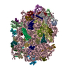
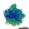
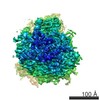
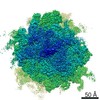
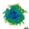
- EMDB-8343: Cryo-EM structure of the Leishmania donovani 80S ribosome at 2.9 ... -
+ Open data

- Basic information
| Entry | Database: EMDB / ID: EMD-8343 | |||||||||||||||||||||
|---|---|---|---|---|---|---|---|---|---|---|---|---|---|---|---|---|---|---|---|---|---|---|
| Title | Cryo-EM structure of the Leishmania donovani 80S ribosome at 2.9 Angstrom resolution | |||||||||||||||||||||
 Map data Map data | Leishmania donovani 80S ribosome | |||||||||||||||||||||
 Sample Sample |
| |||||||||||||||||||||
| Function / homology |  Function and homology information Function and homology informationtranslation regulator activity / maturation of LSU-rRNA from tricistronic rRNA transcript (SSU-rRNA, 5.8S rRNA, LSU-rRNA) / cytosolic ribosome /  ribosomal small subunit assembly / cytosolic small ribosomal subunit / ribosome binding / large ribosomal subunit / small ribosomal subunit / 5S rRNA binding / cytosolic large ribosomal subunit ...translation regulator activity / maturation of LSU-rRNA from tricistronic rRNA transcript (SSU-rRNA, 5.8S rRNA, LSU-rRNA) / cytosolic ribosome / ribosomal small subunit assembly / cytosolic small ribosomal subunit / ribosome binding / large ribosomal subunit / small ribosomal subunit / 5S rRNA binding / cytosolic large ribosomal subunit / rRNA binding / ribosome / structural constituent of ribosome / translation / ribonucleoprotein complex / mRNA binding / nucleolus / RNA binding / zinc ion binding / membrane / metal ion binding / nucleus / cytoplasm ribosomal small subunit assembly / cytosolic small ribosomal subunit / ribosome binding / large ribosomal subunit / small ribosomal subunit / 5S rRNA binding / cytosolic large ribosomal subunit ...translation regulator activity / maturation of LSU-rRNA from tricistronic rRNA transcript (SSU-rRNA, 5.8S rRNA, LSU-rRNA) / cytosolic ribosome / ribosomal small subunit assembly / cytosolic small ribosomal subunit / ribosome binding / large ribosomal subunit / small ribosomal subunit / 5S rRNA binding / cytosolic large ribosomal subunit / rRNA binding / ribosome / structural constituent of ribosome / translation / ribonucleoprotein complex / mRNA binding / nucleolus / RNA binding / zinc ion binding / membrane / metal ion binding / nucleus / cytoplasmSimilarity search - Function | |||||||||||||||||||||
| Biological species |  Leishmania donovani (eukaryote) Leishmania donovani (eukaryote) | |||||||||||||||||||||
| Method | single particle reconstruction / cryo EM / Resolution: 2.9 Å | |||||||||||||||||||||
 Authors Authors | Zhang X / Lai M / Zhou ZH | |||||||||||||||||||||
| Funding support |  United States, 6 items United States, 6 items
| |||||||||||||||||||||
 Citation Citation | Journal: Nat Commun / Year: 2016 Title: Structures and stabilization of kinetoplastid-specific split rRNAs revealed by comparing leishmanial and human ribosomes. Authors: Xing Zhang / Mason Lai / Winston Chang / Iris Yu / Ke Ding / Jan Mrazek / Hwee L Ng / Otto O Yang / Dmitri A Maslov / Z Hong Zhou /  Abstract: The recent success in ribosome structure determination by cryoEM has opened the door to defining structural differences between ribosomes of pathogenic organisms and humans and to understand ribosome- ...The recent success in ribosome structure determination by cryoEM has opened the door to defining structural differences between ribosomes of pathogenic organisms and humans and to understand ribosome-targeting antibiotics. Here, by direct electron-counting cryoEM, we have determined the structures of the Leishmania donovani and human ribosomes at 2.9 Å and 3.6 Å, respectively. Our structure of the leishmanial ribosome elucidates the organization of the six fragments of its large subunit rRNA (as opposed to a single 28S rRNA in most eukaryotes, including humans) and reveals atomic details of a unique 20 amino acid extension of the uL13 protein that pins down the ends of three of the rRNA fragments. The structure also fashions many large rRNA expansion segments. Direct comparison of our human and leishmanial ribosome structures at the decoding A-site sheds light on how the bacterial ribosome-targeting drug paromomycin selectively inhibits the eukaryotic L. donovani, but not human, ribosome. | |||||||||||||||||||||
| History |
|
- Structure visualization
| Movie |
Movie viewer |
|---|---|
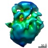
| Structure viewer | EM map: SurfViewMolmilJmol/JSmol |
| Supplemental images |

- Downloads & links
-EMDB archive
| Map data | emd_8343.map.gz | 212.9 MB | EMDB map data format | |
|---|---|---|---|---|
| Header (meta data) | emd-8343-v30.xmlemd-8343.xml | 9.9 KB 9.9 KB | Display Display | EMDB header |
| Images | 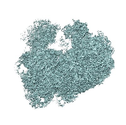 emd_8343.png emd_8343.png | 181 KB | ||
| Archive directory |  http://ftp.pdbj.org/pub/emdb/structures/EMD-8343ftp://ftp.pdbj.org/pub/emdb/structures/EMD-8343 http://ftp.pdbj.org/pub/emdb/structures/EMD-8343ftp://ftp.pdbj.org/pub/emdb/structures/EMD-8343 | HTTPS FTP |
-Related structure data
| Related structure data |  5osgM  5t2aMC 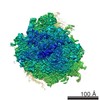 8345C  5t2cC C: citing same article ( M: atomic model generated by this map |
|---|---|
| Similar structure data |
-Links
| EMDB pages | EMDB (EBI/PDBe) / EMDataResource |
|---|---|
| Related items in Molecule of the Month |


-Map
| File | Download / File: emd_8343.map.gz / Format: CCP4 / Size: 244.1 MB / Type: IMAGE STORED AS FLOATING POINT NUMBER (4 BYTES) | ||||||||||||||||||||||||||||||||||||||||||||||||||||||||||||||||||||
|---|---|---|---|---|---|---|---|---|---|---|---|---|---|---|---|---|---|---|---|---|---|---|---|---|---|---|---|---|---|---|---|---|---|---|---|---|---|---|---|---|---|---|---|---|---|---|---|---|---|---|---|---|---|---|---|---|---|---|---|---|---|---|---|---|---|---|---|---|---|
| Annotation | Leishmania donovani 80S ribosome | ||||||||||||||||||||||||||||||||||||||||||||||||||||||||||||||||||||
| Voxel size | X=Y=Z: 0.7933 Å | ||||||||||||||||||||||||||||||||||||||||||||||||||||||||||||||||||||
| Density |
| ||||||||||||||||||||||||||||||||||||||||||||||||||||||||||||||||||||
| Symmetry | Space group: 1 | ||||||||||||||||||||||||||||||||||||||||||||||||||||||||||||||||||||
| Details | EMDB XML:
CCP4 map header:
| ||||||||||||||||||||||||||||||||||||||||||||||||||||||||||||||||||||
-Supplemental data
- Sample components
-Entire : 80S ribosome
| Entire | Name: 80S ribosomeEukaryotic ribosome |
|---|---|
| Components |
|
-Supramolecule #1: 80S ribosome
| Supramolecule | Name: 80S ribosome / type: complex / ID: 1 / Parent: 0 |
|---|---|
| Source (natural) | Organism: Leishmania donovani (eukaryote) / Strain: 1S |
-Experimental details
-Structure determination
| Method | cryo EM |
|---|---|
 Processing Processing | single particle reconstruction |
| Aggregation state | particle |
-Sample preparation
| Buffer | pH: 7.6 |
|---|---|
| Vitrification | Cryogen name: ETHANE / Chamber humidity: 100 % |
- Electron microscopy
| Microscope | FEI TITAN KRIOS |
|---|---|
| Electron beam | Acceleration voltage: 300 kV / Electron source: FIELD EMISSION GUN |
| Electron optics | Illumination mode: FLOOD BEAM / Imaging mode: BRIGHT FIELDBright-field microscopy |
| Image recording | Film or detector model: GATAN K2 SUMMIT (4k x 4k) / Detector mode: COUNTING / Average electron dose: 20.0 e/Å2 |
| Experimental equipment |  Model: Titan Krios / Image courtesy: FEI Company |
-Image processing
| CTF correction | Software - Name: CTFFIND |
|---|---|
| Startup model | Type of model: EMDB MAP EMDB ID: |
| Initial angle assignment | Type: NOT APPLICABLE |
| Final angle assignment | Type: OTHER |
| Final reconstruction | Resolution.type: BY AUTHOR / Resolution: 2.9 Å / Resolution method: FSC 0.143 CUT-OFF / Number images used: 213108 |