- EMDB-8235: Negative stain structure of Vps15/Vps34 complex -
+
Open data
ID or keywords:
Loading...
-
Basic information
Entry
Database: EMDB / ID: EMD-8235
Title
Negative stain structure of Vps15/Vps34 complex
Map data
Vps15/34 complex
Sample
Complex: Vps15/34
Protein or peptide: Phosphatidylinositol 3-kinase VPS34
Protein or peptide: Serine/threonine-protein kinase VPS15
Function / homology
Function and homology information
Synthesis of PIPs at the late endosome membrane / Synthesis of PIPs at the early endosome membrane / RHO GTPases Activate NADPH Oxidases / autophagy of peroxisome / nucleus-vacuole junction / Synthesis of PIPs at the Golgi membrane / vacuole-isolation membrane contact site / vacuole inheritance / phosphatidylinositol 3-kinase complex, class III, type II / phosphatidylinositol 3-kinase complex, class III, type I ...Synthesis of PIPs at the late endosome membrane / Synthesis of PIPs at the early endosome membrane / RHO GTPases Activate NADPH Oxidases / autophagy of peroxisome / nucleus-vacuole junction / Synthesis of PIPs at the Golgi membrane / vacuole-isolation membrane contact site / vacuole inheritance / phosphatidylinositol 3-kinase complex, class III, type II / phosphatidylinositol 3-kinase complex, class III, type I / Macroautophagy / protein retention in Golgi apparatus / pexophagy / phagophore assembly site membrane / protein targeting to vacuole / late endosome to vacuole transport / fungal-type vacuole membrane / phagophore assembly site / phosphatidylinositol-mediated signaling / phosphatidylinositol 3-kinase / phosphatidylinositol-3-phosphate biosynthetic process / 1-phosphatidylinositol-3-kinase activity / phosphatidylinositol phosphate biosynthetic process / autophagosome assembly / ubiquitin binding / positive regulation of transcription elongation by RNA polymerase II / macroautophagy / autophagy / peroxisome / endocytosis / protein transport / late endosome / endosome membrane / non-specific serine/threonine protein kinase / endosome / protein kinase activity / phosphorylation / Golgi membrane / protein serine kinase activity / protein serine/threonine kinase activity / mitochondrion / ATP binding / membrane / cytosol / cytoplasm Similarity search - Function
Journal: Autophagy / Year: 2016 Title: Characterization of Atg38 and NRBF2, a fifth subunit of the autophagic Vps34/PIK3C3 complex. Authors: Yohei Ohashi / Nicolas Soler / Miguel García Ortegón / Lufei Zhang / Marie L Kirsten / Olga Perisic / Glenn R Masson / John E Burke / Arjen J Jakobi / Apostolos A Apostolakis / Christopher ...Authors: Yohei Ohashi / Nicolas Soler / Miguel García Ortegón / Lufei Zhang / Marie L Kirsten / Olga Perisic / Glenn R Masson / John E Burke / Arjen J Jakobi / Apostolos A Apostolakis / Christopher M Johnson / Maki Ohashi / Nicholas T Ktistakis / Carsten Sachse / Roger L Williams / Abstract: The phosphatidylinositol 3-kinase Vps34 is part of several protein complexes. The structural organization of heterotetrameric complexes is starting to emerge, but little is known about organization ...The phosphatidylinositol 3-kinase Vps34 is part of several protein complexes. The structural organization of heterotetrameric complexes is starting to emerge, but little is known about organization of additional accessory subunits that interact with these assemblies. Combining hydrogen-deuterium exchange mass spectrometry (HDX-MS), X-ray crystallography and electron microscopy (EM), we have characterized Atg38 and its human ortholog NRBF2, accessory components of complex I consisting of Vps15-Vps34-Vps30/Atg6-Atg14 (yeast) and PIK3R4/VPS15-PIK3C3/VPS34-BECN1/Beclin 1-ATG14 (human). HDX-MS shows that Atg38 binds the Vps30-Atg14 subcomplex of complex I, using mainly its N-terminal MIT domain and bridges the coiled-coil I regions of Atg14 and Vps30 in the base of complex I. The Atg38 C-terminal domain is important for localization to the phagophore assembly site (PAS) and homodimerization. Our 2.2 Å resolution crystal structure of the Atg38 C-terminal homodimerization domain shows 2 segments of α-helices assembling into a mushroom-like asymmetric homodimer with a 4-helix cap and a parallel coiled-coil stalk. One Atg38 homodimer engages a single complex I. This is in sharp contrast to human NRBF2, which also forms a homodimer, but this homodimer can bridge 2 complex I assemblies.
History
Deposition
Jun 4, 2016
-
Header (metadata) release
Jun 22, 2016
-
Map release
Oct 5, 2016
-
Update
Nov 13, 2019
-
Current status
Nov 13, 2019
Processing site: PDBe / Status: Released
-
Structure visualization
Movie
Surface view with section colored by density value
In the structure databanks used in Yorodumi, some data are registered as the other names, "COVID-19 virus" and "2019-nCoV". Here are the details of the virus and the list of structure data.
Jan 31, 2019. EMDB accession codes are about to change! (news from PDBe EMDB page)
EMDB accession codes are about to change! (news from PDBe EMDB page)
The allocation of 4 digits for EMDB accession codes will soon come to an end. Whilst these codes will remain in use, new EMDB accession codes will include an additional digit and will expand incrementally as the available range of codes is exhausted. The current 4-digit format prefixed with “EMD-” (i.e. EMD-XXXX) will advance to a 5-digit format (i.e. EMD-XXXXX), and so on. It is currently estimated that the 4-digit codes will be depleted around Spring 2019, at which point the 5-digit format will come into force.
The EM Navigator/Yorodumi systems omit the EMD- prefix.
Related info.:Q: What is EMD? / ID/Accession-code notation in Yorodumi/EM Navigator
Yorodumi is a browser for structure data from EMDB, PDB, SASBDB, etc.
This page is also the successor to EM Navigator detail page, and also detail information page/front-end page for Omokage search.
The word "yorodu" (or yorozu) is an old Japanese word meaning "ten thousand". "mi" (miru) is to see.
Related info.:EMDB / PDB / SASBDB / Comparison of 3 databanks / Yorodumi Search / Aug 31, 2016. New EM Navigator & Yorodumi / Yorodumi Papers / Jmol/JSmol / Function and homology information / Changes in new EM Navigator and Yorodumi
 Movie
Movie Controller


 Open data
 Basic information
 Map data
 Sample
 Function and homology information
 Macroautophagy / protein retention in Golgi apparatus / pexophagy / phagophore assembly site membrane / protein targeting to vacuole / late endosome to vacuole transport / fungal-type vacuole membrane / phagophore assembly site / phosphatidylinositol-mediated signaling /
 Authors
 Citation

 Structure visualization
 Downloads & links
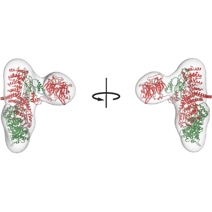 emd_8235.png
 http://ftp.pdbj.org/pub/emdb/structures/EMD-8235


 Z (Sec.)
 Y (Row.)
 X (Col.)


 Sample components
 Processing
 Electron microscopy
