 Movie
Movie Controller

[English] 日本語
 Yorodumi
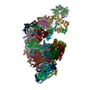
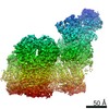
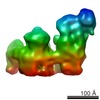
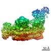
- EMDB-8130: Architecture of ovine respiratory supercomplex (I-III2-IV) confor... -
+ Open data

- Basic information
| Entry | Database: EMDB / ID: EMD-8130 | |||||||||
|---|---|---|---|---|---|---|---|---|---|---|
| Title | Architecture of ovine respiratory supercomplex (I-III2-IV) conformation tight | |||||||||
 Map data Map data | None | |||||||||
 Sample Sample |
| |||||||||
| Function / homology |  Function and homology information Function and homology informationrespiratory chain complex III / respiratory chain complex IV assembly / respiratory chain complex IV / aerobic electron transport chain / mitochondrial respiratory chain complex III / mitochondrial respiratory chain complex IV / mitochondrial respirasome /  cytochrome-c oxidase / quinol-cytochrome-c reductase / ubiquinol-cytochrome-c reductase activity ...respiratory chain complex III / respiratory chain complex IV assembly / respiratory chain complex IV / aerobic electron transport chain / mitochondrial respiratory chain complex III / mitochondrial respiratory chain complex IV / mitochondrial respirasome / cytochrome-c oxidase / quinol-cytochrome-c reductase / ubiquinol-cytochrome-c reductase activity / oxidative phosphorylation / mitochondrial electron transport, cytochrome c to oxygen / cytochrome-c oxidase activity / mitochondrial electron transport, ubiquinol to cytochrome c / respirasome / respiratory electron transport chain / 2 iron, 2 sulfur cluster binding / metalloendopeptidase activity / mitochondrial inner membrane / electron transfer activity / membrane => GO:0016020 / copper ion binding / ubiquitin protein ligase binding / heme binding / proteolysis / nucleoplasm / metal ion binding cytochrome-c oxidase / quinol-cytochrome-c reductase / ubiquinol-cytochrome-c reductase activity ...respiratory chain complex III / respiratory chain complex IV assembly / respiratory chain complex IV / aerobic electron transport chain / mitochondrial respiratory chain complex III / mitochondrial respiratory chain complex IV / mitochondrial respirasome / cytochrome-c oxidase / quinol-cytochrome-c reductase / ubiquinol-cytochrome-c reductase activity / oxidative phosphorylation / mitochondrial electron transport, cytochrome c to oxygen / cytochrome-c oxidase activity / mitochondrial electron transport, ubiquinol to cytochrome c / respirasome / respiratory electron transport chain / 2 iron, 2 sulfur cluster binding / metalloendopeptidase activity / mitochondrial inner membrane / electron transfer activity / membrane => GO:0016020 / copper ion binding / ubiquitin protein ligase binding / heme binding / proteolysis / nucleoplasm / metal ion bindingSimilarity search - Function | |||||||||
| Biological species |  Ovis aries (sheep) Ovis aries (sheep) | |||||||||
| Method | single particle reconstruction / cryo EM / Resolution: 5.8 Å | |||||||||
 Authors Authors | Sazanov LA / Letts JA / Fiedorczuk K | |||||||||
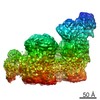
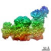
 Citation Citation | Journal: Nature / Year: 2016 Title: The architecture of respiratory supercomplexes. Authors: James A Letts / Karol Fiedorczuk / Leonid A Sazanov /   Abstract: Mitochondrial electron transport chain complexes are organized into supercomplexes responsible for carrying out cellular respiration. Here we present three architectures of mammalian (ovine) ...Mitochondrial electron transport chain complexes are organized into supercomplexes responsible for carrying out cellular respiration. Here we present three architectures of mammalian (ovine) supercomplexes determined by cryo-electron microscopy. We identify two distinct arrangements of supercomplex CICIIICIV (the respirasome)-a major 'tight' form and a minor 'loose' form (resolved at the resolution of 5.8 Å and 6.7 Å, respectively), which may represent different stages in supercomplex assembly or disassembly. We have also determined an architecture of supercomplex CICIII at 7.8 Å resolution. All observed density can be attributed to the known 80 subunits of the individual complexes, including 132 transmembrane helices. The individual complexes form tight interactions that vary between the architectures, with complex IV subunit COX7a switching contact from complex III to complex I. The arrangement of active sites within the supercomplex may help control reactive oxygen species production. To our knowledge, these are the first complete architectures of the dominant, physiologically relevant state of the electron transport chain. | |||||||||
| History |
|
- Structure visualization
| Movie |
Movie viewer |
|---|---|
| Structure viewer | EM map: SurfViewMolmilJmol/JSmol |
| Supplemental images |

- Downloads & links
-EMDB archive
| Map data | emd_8130.map.gz | 436.9 MB | EMDB map data format | |
|---|---|---|---|---|
| Header (meta data) | emd-8130-v30.xmlemd-8130.xml | 31.6 KB 31.6 KB | Display Display | EMDB header |
| FSC (resolution estimation) | emd_8130_fsc.xml | 17.1 KB | Display | FSC data file |
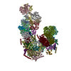
| Images | 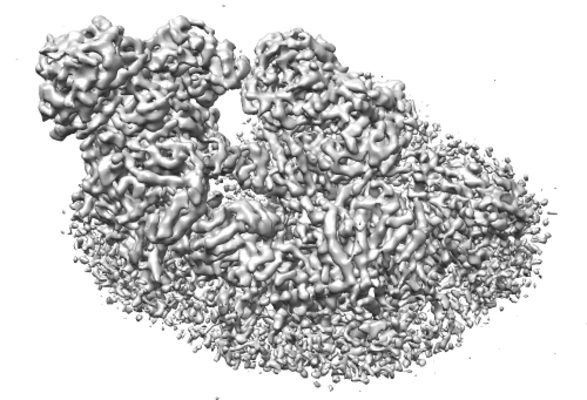 emd_8130.png emd_8130.png | 126.3 KB | ||
| Archive directory |  http://ftp.pdbj.org/pub/emdb/structures/EMD-8130ftp://ftp.pdbj.org/pub/emdb/structures/EMD-8130 http://ftp.pdbj.org/pub/emdb/structures/EMD-8130ftp://ftp.pdbj.org/pub/emdb/structures/EMD-8130 | HTTPS FTP |
-Related structure data
| Related structure data | 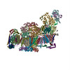 5j4zMC  8128C  8129C 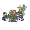 5j7yC 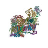 5j8kC C: citing same article ( M: atomic model generated by this map |
|---|---|
| Similar structure data |
-Links
| EMDB pages | EMDB (EBI/PDBe) / EMDataResource |
|---|---|
| Related items in Molecule of the Month |


-Map
| File | Download / File: emd_8130.map.gz / Format: CCP4 / Size: 465.5 MB / Type: IMAGE STORED AS FLOATING POINT NUMBER (4 BYTES) | ||||||||||||||||||||||||||||||||||||||||||||||||||||||||||||
|---|---|---|---|---|---|---|---|---|---|---|---|---|---|---|---|---|---|---|---|---|---|---|---|---|---|---|---|---|---|---|---|---|---|---|---|---|---|---|---|---|---|---|---|---|---|---|---|---|---|---|---|---|---|---|---|---|---|---|---|---|---|
| Annotation | None | ||||||||||||||||||||||||||||||||||||||||||||||||||||||||||||
| Projections & slices | Image control
Images are generated by Spider. | ||||||||||||||||||||||||||||||||||||||||||||||||||||||||||||
| Voxel size | X=Y=Z: 1.72 Å | ||||||||||||||||||||||||||||||||||||||||||||||||||||||||||||
| Density |
| ||||||||||||||||||||||||||||||||||||||||||||||||||||||||||||
| Symmetry | Space group: 1 | ||||||||||||||||||||||||||||||||||||||||||||||||||||||||||||
| Details | EMDB XML:
CCP4 map header:
| ||||||||||||||||||||||||||||||||||||||||||||||||||||||||||||
 Z (Sec.)
 Y (Row.)
 X (Col.)


-Supplemental data
- Sample components
-Entire : Ovine respiratory supercomplex (I-III2-IV), conformation tight
| Entire | Name: Ovine respiratory supercomplex (I-III2-IV), conformation tight |
|---|---|
| Components |
|
-Supramolecule #1: Ovine respiratory supercomplex (I-III2-IV), conformation tight
| Supramolecule | Name: Ovine respiratory supercomplex (I-III2-IV), conformation tight type: complex / ID: 1 / Parent: 0 / Macromolecule list: #1-#68 |
|---|---|
| Source (natural) | Organism: Ovis aries (sheep) |
| Molecular weight | Experimental: 1.71 MDa |
-Supramolecule #2: Respiratory complex IV
| Supramolecule | Name: Respiratory complex IV / type: complex / ID: 2 / Parent: 1 / Macromolecule list: #1-#13 / Details: Monomer |
|---|---|
| Source (natural) | Organism: Ovis aries (sheep) |
| Molecular weight | Theoretical: 220 KDa |
-Supramolecule #3: Respiratory complex III
| Supramolecule | Name: Respiratory complex III / type: complex / ID: 3 / Parent: 1 / Macromolecule list: #14-#24 / Details: Dimer |
|---|---|
| Source (natural) | Organism: Ovis aries (sheep) |
| Molecular weight | Theoretical: 490 KDa |
-Supramolecule #4: Respiratory complex I
| Supramolecule | Name: Respiratory complex I / type: complex / ID: 4 / Parent: 1 / Macromolecule list: #25-#68 / Details: Monomer |
|---|---|
| Source (natural) | Organism: Ovis aries (sheep) |
| Molecular weight | Theoretical: 1.0 MDa |
-Experimental details
-Structure determination
| Method | cryo EM |
|---|---|
 Processing Processing | single particle reconstruction |
| Aggregation state | particle |
-Sample preparation
| Concentration | 2.0 mg/mL | ||||||||||||
|---|---|---|---|---|---|---|---|---|---|---|---|---|---|
| Buffer | pH: 7.7 Component:
| ||||||||||||
| Grid | Model: Quantifoil R0.6/1 / Material: COPPER / Mesh: 300 / Pretreatment - Type: GLOW DISCHARGE / Pretreatment - Atmosphere: AIR | ||||||||||||
| Vitrification | Cryogen name: ETHANE / Chamber humidity: 100 % / Chamber temperature: 277 K / Instrument: FEI VITROBOT MARK IV / Details: Blotted 32 seconds. | ||||||||||||
| Details | Single particles dispersed in digitonin |
- Electron microscopy
| Microscope | FEI TITAN KRIOS |
|---|---|
| Electron beam | Acceleration voltage: 300 kV / Electron source: FIELD EMISSION GUN |
| Electron optics | C2 aperture diameter: 70.0 µm / Calibrated magnification: 81395 / Illumination mode: FLOOD BEAM / Imaging mode: BRIGHT FIELDBright-field microscopy / Cs: 2.7 mm / Nominal defocus max: 5.0 µm / Nominal defocus min: 1.0 µm / Nominal magnification: 47000 |
| Sample stage | Specimen holder model: FEI TITAN KRIOS AUTOGRID HOLDER / Cooling holder cryogen: NITROGEN |
| Image recording | Film or detector model: FEI FALCON II (4k x 4k) / Detector mode: INTEGRATING / Digitization - Dimensions - Width: 4096 pixel / Digitization - Dimensions - Height: 4096 pixel / Digitization - Sampling interval: 14.0 µm / Digitization - Frames/image: 1-32 / Number grids imaged: 3 / Number real images: 1608 / Average exposure time: 4.0 sec. / Average electron dose: 34.0 e/Å2 Details: Images were collected in movie mode with 17 frames per second. |
| Experimental equipment |  Model: Titan Krios / Image courtesy: FEI Company |
-Image processing
| Particle selection | Number selected: 67650 |
|---|---|
| CTF correction | Software - Name: Gctf |
| Initial angle assignment | Type: PROJECTION MATCHING |
| Final angle assignment | Type: PROJECTION MATCHING |
| Final reconstruction | Number classes used: 1 / Resolution.type: BY AUTHOR / Resolution: 5.8 Å / Resolution method: FSC 0.143 CUT-OFF / Software - Name: RELION (ver. 1.4) / Number images used: 18379 |
| FSC plot (resolution estimation) |  |
-Atomic model buiding 1
| Refinement | Space: REAL / Protocol: BACKBONE TRACE |
|---|---|
| Output model | PDB-5j4z: |
