 Movie
Movie Controller

[English] 日本語
 Yorodumi
- EMDB-8096: The Architecture of the Cytoplasmic Region of Type III Secretion ... -
+ Open data

- Basic information
| Entry | Database: EMDB / ID: EMD-8096 | |||||||||
|---|---|---|---|---|---|---|---|---|---|---|
| Title | The Architecture of the Cytoplasmic Region of Type III Secretion Systems Type three secretion system Type three secretion system | |||||||||
 Map data Map data | None | |||||||||
 Sample Sample |
| |||||||||
| Biological species |  Shigella flexneri (bacteria) Shigella flexneri (bacteria) | |||||||||
| Method | subtomogram averaging / cryo EM / Resolution: 50.0 Å | |||||||||
 Authors Authors | Makino F / Shen D / Kajimura N / Kawamoto A / Pissaridou P / Oswin H / Pain M / Murillo I / Namba K / Blocker AJ | |||||||||
 Citation Citation | Journal: Sci Rep / Year: 2016 Title: The Architecture of the Cytoplasmic Region of Type III Secretion Systems. Authors: Fumiaki Makino / Dakang Shen / Naoko Kajimura / Akihiro Kawamoto / Panayiota Pissaridou / Henry Oswin / Maria Pain / Isabel Murillo / Keiichi Namba / Ariel J Blocker /   Abstract: Type III secretion systems (T3SSs) are essential devices in the virulence of many Gram-negative bacterial pathogens. They mediate injection of protein effectors of virulence from bacteria into ...Type III secretion systems (T3SSs) are essential devices in the virulence of many Gram-negative bacterial pathogens. They mediate injection of protein effectors of virulence from bacteria into eukaryotic host cells to manipulate them during infection. T3SSs involved in virulence (vT3SSs) are evolutionarily related to bacterial flagellar protein export apparatuses (fT3SSs), which are essential for flagellar assembly and cell motility. The structure of the external and transmembrane parts of both fT3SS and vT3SS is increasingly well-defined. However, the arrangement of their cytoplasmic and inner membrane export apparatuses is much less clear. Here we compare the architecture of the cytoplasmic regions of the vT3SSs of Shigella flexneri and the vT3SS and fT3SS of Salmonella enterica serovar Typhimurium at ~5 and ~4 nm resolution using electron cryotomography and subtomogram averaging. We show that the cytoplasmic regions of vT3SSs display conserved six-fold symmetric features including pods, linkers and an ATPase complex, while fT3SSs probably only display six-fold symmetry in their ATPase region. We also identify other morphological differences between vT3SSs and fT3SSs, such as relative disposition of their inner membrane-attached export platform, C-ring/pods and ATPase complex. Finally, using classification, we find that both types of apparatuses can loose elements of their cytoplasmic region, which may therefore be dynamic. | |||||||||
| History |
|
- Structure visualization
| Movie |
 Movie viewer Movie viewer |
|---|---|
| Structure viewer | EM map: SurfViewMolmilJmol/JSmol |
| Supplemental images |

- Downloads & links
-EMDB archive
| Map data | emd_8096.map.gz | 1.5 MB | EMDB map data format | |
|---|---|---|---|---|
| Header (meta data) | emd-8096-v30.xmlemd-8096.xml | 11.2 KB 11.2 KB | Display Display | EMDB header |
| Images | 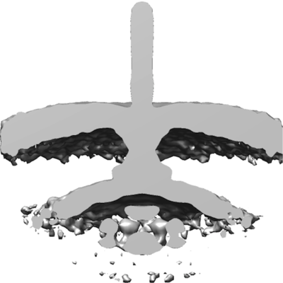 emd_8096.png emd_8096.png | 62 KB | ||
| Archive directory |  http://ftp.pdbj.org/pub/emdb/structures/EMD-8096ftp://ftp.pdbj.org/pub/emdb/structures/EMD-8096 http://ftp.pdbj.org/pub/emdb/structures/EMD-8096ftp://ftp.pdbj.org/pub/emdb/structures/EMD-8096 | HTTPS FTP |
-Related structure data


-Links
| EMDB pages | EMDB (EBI/PDBe) / EMDataResource |
|---|
-Map
| File | Download / File: emd_8096.map.gz / Format: CCP4 / Size: 3.8 MB / Type: IMAGE STORED AS FLOATING POINT NUMBER (4 BYTES) | ||||||||||||||||||||||||||||||||||||||||||||||||||||||||||||||||||||
|---|---|---|---|---|---|---|---|---|---|---|---|---|---|---|---|---|---|---|---|---|---|---|---|---|---|---|---|---|---|---|---|---|---|---|---|---|---|---|---|---|---|---|---|---|---|---|---|---|---|---|---|---|---|---|---|---|---|---|---|---|---|---|---|---|---|---|---|---|---|
| Annotation | None | ||||||||||||||||||||||||||||||||||||||||||||||||||||||||||||||||||||
| Projections & slices | Image control
Images are generated by Spider. | ||||||||||||||||||||||||||||||||||||||||||||||||||||||||||||||||||||
| Voxel size | X=Y=Z: 5.7 Å | ||||||||||||||||||||||||||||||||||||||||||||||||||||||||||||||||||||
| Density |
| ||||||||||||||||||||||||||||||||||||||||||||||||||||||||||||||||||||
| Symmetry | Space group: 1 | ||||||||||||||||||||||||||||||||||||||||||||||||||||||||||||||||||||
| Details | EMDB XML:
CCP4 map header:
| ||||||||||||||||||||||||||||||||||||||||||||||||||||||||||||||||||||
 Z (Sec.)
 Y (Row.)
 X (Col.)


-Supplemental data
- Sample components
-Entire : The needle complex of Shigella flexneri
| Entire | Name: The needle complex of Shigella flexneri |
|---|---|
| Components |
|
-Supramolecule #1: The needle complex of Shigella flexneri
| Supramolecule | Name: The needle complex of Shigella flexneri / type: complex / ID: 1 / Parent: 0 |
|---|---|
| Source (natural) | Organism: Shigella flexneri (bacteria) |
| Recombinant expression | Organism: Shigella flexneri (bacteria) / Recombinant strain: WT for minicells / Recombinant plasmid: pBAD24 Salmonella ftsZ |
-Experimental details
-Structure determination
| Method | cryo EM |
|---|---|
 Processing Processing | subtomogram averaging |
| Aggregation state | 3D array |
-Sample preparation
| Buffer | pH: 7 |
|---|---|
| Grid | Model: Quantifoil R0.6/1.0 / Material: MOLYBDENUM / Mesh: 200 / Support film - Material: CARBON / Support film - topology: HOLEY / Pretreatment - Type: GLOW DISCHARGE |
| Vitrification | Cryogen name: ETHANE / Chamber humidity: 100 % / Chamber temperature: 277 K / Instrument: FEI VITROBOT MARK III |
- Electron microscopy
| Microscope | FEI TITAN KRIOS |
|---|---|
| Electron beam | Acceleration voltage: 300 kV / Electron source: FIELD EMISSION GUN |
| Electron optics | Calibrated magnification: 49030 / Illumination mode: FLOOD BEAM / Imaging mode: BRIGHT FIELDBright-field microscopy / Cs: 2.7 mm / Nominal defocus max: 10.0 µm / Nominal defocus min: 5.0 µm / Nominal magnification: 29000 |
| Sample stage | Specimen holder model: FEI TITAN KRIOS AUTOGRID HOLDER / Cooling holder cryogen: NITROGEN |
| Temperature | Min: 80.0 K / Max: 80.0 K |
| Image recording | Film or detector model: FEI FALCON II (4k x 4k) / Detector mode: INTEGRATING / Average exposure time: 1.0 sec. / Average electron dose: 3.0 e/Å2 |
| Experimental equipment |  Model: Titan Krios / Image courtesy: FEI Company |
-Image processing
| Extraction | Number tomograms: 100 / Number images used: 265 / Method: manual / Software - Name: IMOD |
|---|---|
| CTF correction | Software - Name: TOMOCTF |
| Final 3D classification | Number classes: 4 / Avg.num./class: 70 / Software - Name: Dynamo |
| Final angle assignment | Type: ANGULAR RECONSTITUTION / Software - Name: Dynamo |
| Final reconstruction | Applied symmetry - Point group: C6 (6 fold cyclic) / Algorithm: SIMULTANEOUS ITERATIVE (SIRT) / Resolution.type: BY AUTHOR / Resolution: 50.0 Å / Resolution method: FSC 0.143 CUT-OFF / Software - Name: Dynamo / Number subtomograms used: 265 |
