 Movie
Movie Controller


+ Open data

- Basic information
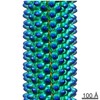
| Entry | Database: EMDB / ID: EMD-8065 | ||||||||||||
|---|---|---|---|---|---|---|---|---|---|---|---|---|---|
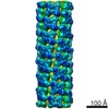
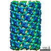
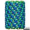
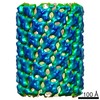
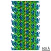
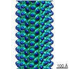
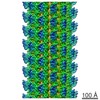
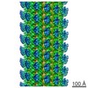
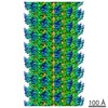
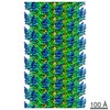
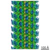
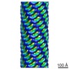
| Title | Soybean agglutinin(SBA) microtube | ||||||||||||
 Map data Map data | None | ||||||||||||
 Sample Sample |
| ||||||||||||
| Biological species |  | ||||||||||||
| Method | helical reconstruction / cryo EM / Resolution: 7.8 Å | ||||||||||||
 Authors Authors | Zhang X / Li X / Liu C | ||||||||||||
| Funding support |  China, 3 items China, 3 items
| ||||||||||||
 Citation Citation | Journal: J Am Chem Soc / Year: 2019 Title: Chemically Controlled Helical Polymorphism in Protein Tubes by Selective Modulation of Supramolecular Interactions. Authors: Zhen Li / Shuyu Chen / Chendi Gao / Zhiwei Yang / Kuo-Chih Shih / Zdravko Kochovski / Guang Yang / Lu Gou / Mu-Ping Nieh / Ming Jiang / Lei Zhang / Guosong Chen /   Abstract: Polymorphism has been the subject of investigation across different research disciplines. In biology, polymorphism could be interpreted in such a way that discrete biomacromolecules can adopt ...Polymorphism has been the subject of investigation across different research disciplines. In biology, polymorphism could be interpreted in such a way that discrete biomacromolecules can adopt diversiform specific conformations/packing arrangement, and this polymorph-dependent property is essential for many biochemical processes. For example, bacterial flagellar filament, composed of flagellin, switches between different supercoiled state allowing the bacteria to swim and tumble. However, in artificial supramolecular systems, it is often challenging to achieve polymorph control and prediction, and in most cases, two or more concomitant polymorphs of similar formation energies coexist. Here, we show that a tetrameric protein with properly oriented binding sites on its surface can arrange into diverse protein tubes with distinct helical parameters by adding specifically designed inducing ligands. We examined several parameters of the ligand that would influence the protein tube formation and found that the flexibility of the ligand linker and the dimerization pose of the ligand complex is critical for the successful production of the tubes and eventually influence the specific helical polymorphs of the formed tubes. A surface lattice accommodation model was further developed to rationalize the geometrical relationship between each helical tube type. Molecular simulation was used to elucidate the interactions between ligands and SBA and molecular basis for polymorphic switching of the protein tubes. Moreover, the kinetics of structural formation was studied and the ligand design was found that can affect the kinetics of the protein polymerization pathway. In short, our designed protein tubes serves as an enlightening system for understanding how a protein polymer composed of a single protein switches among different helical states. | ||||||||||||
| History |
|
- Structure visualization
| Movie |
 Movie viewer Movie viewer |
|---|---|
| Structure viewer | EM map: SurfViewMolmilJmol/JSmol |
| Supplemental images |

- Downloads & links
-EMDB archive
| Map data | emd_8065.map.gz | 8.6 MB | EMDB map data format | |
|---|---|---|---|---|
| Header (meta data) | emd-8065-v30.xmlemd-8065.xml | 11.5 KB 11.5 KB | Display Display | EMDB header |
| Images |  emd_8065.png emd_8065.png | 54.6 KB | ||
| Archive directory |  http://ftp.pdbj.org/pub/emdb/structures/EMD-8065ftp://ftp.pdbj.org/pub/emdb/structures/EMD-8065 http://ftp.pdbj.org/pub/emdb/structures/EMD-8065ftp://ftp.pdbj.org/pub/emdb/structures/EMD-8065 | HTTPS FTP |
-Validation report
| Summary document | emd_8065_validation.pdf.gz | 79.2 KB | Display | EMDB validaton report |
|---|---|---|---|---|
| Full document | emd_8065_full_validation.pdf.gz | 78.3 KB | Display | |
| Data in XML | emd_8065_validation.xml.gz | 493 B | Display | |
| Arichive directory | https://ftp.pdbj.org/pub/emdb/validation_reports/EMD-8065ftp://ftp.pdbj.org/pub/emdb/validation_reports/EMD-8065 | HTTPS FTP |
-Related structure data


-Links
| EMDB pages | EMDB (EBI/PDBe) / EMDataResource |
|---|
-Map
| File | Download / File: emd_8065.map.gz / Format: CCP4 / Size: 52.7 MB / Type: IMAGE STORED AS FLOATING POINT NUMBER (4 BYTES) | ||||||||||||||||||||||||||||||||||||||||||||||||||||||||||||||||||||
|---|---|---|---|---|---|---|---|---|---|---|---|---|---|---|---|---|---|---|---|---|---|---|---|---|---|---|---|---|---|---|---|---|---|---|---|---|---|---|---|---|---|---|---|---|---|---|---|---|---|---|---|---|---|---|---|---|---|---|---|---|---|---|---|---|---|---|---|---|---|
| Annotation | None | ||||||||||||||||||||||||||||||||||||||||||||||||||||||||||||||||||||
| Voxel size | X=Y=Z: 2.64 Å | ||||||||||||||||||||||||||||||||||||||||||||||||||||||||||||||||||||
| Density |
| ||||||||||||||||||||||||||||||||||||||||||||||||||||||||||||||||||||
| Symmetry | Space group: 1 | ||||||||||||||||||||||||||||||||||||||||||||||||||||||||||||||||||||
| Details | EMDB XML:
CCP4 map header:
| ||||||||||||||||||||||||||||||||||||||||||||||||||||||||||||||||||||
-Supplemental data
- Sample components
-Entire : soybean agglutinin microtube
| Entire | Name: soybean agglutinin microtube |
|---|---|
| Components |
|
-Supramolecule #1: soybean agglutinin microtube
| Supramolecule | Name: soybean agglutinin microtube / type: complex / ID: 1 / Parent: 0 Details: soybean agglutinin tetramer linked by the designed ligands |
|---|---|
| Source (natural) | Organism: |
-Experimental details
-Structure determination
| Method | cryo EM |
|---|---|
 Processing Processing | helical reconstruction |
| Aggregation state | helical array |
-Sample preparation
| Buffer | pH: 7 |
|---|---|
| Grid | Model: Qutantifoil R1.2/1.3 / Material: COPPER / Mesh: 400 / Support film - Material: CARBON / Support film - topology: HOLEY ARRAY / Pretreatment - Type: GLOW DISCHARGE / Pretreatment - Atmosphere: AIR |
| Vitrification | Cryogen name: ETHANE / Chamber humidity: 100 % / Instrument: FEI VITROBOT MARK IV |
- Electron microscopy
| Microscope | FEI TITAN KRIOS |
|---|---|
| Image recording | Film or detector model: GATAN K2 SUMMIT (4k x 4k) / Detector mode: SUPER-RESOLUTION / Digitization - Frames/image: 1-32 / Average exposure time: 8.0 sec. / Average electron dose: 50.0 e/Å2 / Details: Images were collected with 4 frames per second |
| Electron beam | Acceleration voltage: 300 kV / Electron source:  FIELD EMISSION GUN FIELD EMISSION GUN |
| Electron optics | C2 aperture diameter: 50.0 µm / Calibrated defocus max: 2.0 µm / Calibrated defocus min: 1.0 µm / Illumination mode: FLOOD BEAM / Imaging mode: BRIGHT FIELD / Cs: 2.7 mm / Nominal defocus max: 2.0 µm / Nominal defocus min: 1.0 µm / Nominal magnification: 22500 |
| Sample stage | Specimen holder model: FEI TITAN KRIOS AUTOGRID HOLDER / Cooling holder cryogen: NITROGEN |
| Experimental equipment |  Model: Titan Krios / Image courtesy: FEI Company |
-Image processing
| Final reconstruction | Applied symmetry - Helical parameters - Δz: 21.22 Å Applied symmetry - Helical parameters - Δ&Phi: -36.9 ° Applied symmetry - Helical parameters - Axial symmetry: C1 (asymmetric) Resolution.type: BY AUTHOR / Resolution: 7.8 Å / Resolution method: FSC 0.143 CUT-OFF / Software - Name: FREALIGN (ver. 8.09) / Number images used: 63925 |
|---|---|
| CTF correction | Software - Name: CTFFIND (ver. 3) |
| Startup model | Type of model: OTHER / Details: cylinder |
| Final angle assignment | Type: NOT APPLICABLE / Software - Name: FREALIGN Details: Local refinement was performed using FREALIGN mode 1 |
-Atomic model buiding 1
| Refinement | Protocol: RIGID BODY FIT |
|---|
