 Movie
Movie Controller

[English] 日本語
 Yorodumi
- EMDB-6678: CryoEM structure of type II secretion system cap deletion secreti... -
+ Open data

- Basic information
| Entry | Database: EMDB / ID: EMD-6678 | |||||||||
|---|---|---|---|---|---|---|---|---|---|---|
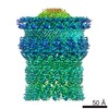
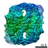
| Title | CryoEM structure of type II secretion system cap deletion secretin GspD in Vibrio cholerae Type II secretion system Type II secretion system | |||||||||
 Map data Map data | VC_GspD_DelCap post-processing map | |||||||||
 Sample Sample |
| |||||||||
| Function / homology |  Function and homology informationprotein secretion by the type II secretion system / type II protein secretion system complex / protein secretion / cell outer membrane / identical protein binding Function and homology informationprotein secretion by the type II secretion system / type II protein secretion system complex / protein secretion / cell outer membrane / identical protein bindingSimilarity search - Function | |||||||||
| Biological species |  Vibrio cholerae O1 biovar El Tor str. N16961 (bacteria) Vibrio cholerae O1 biovar El Tor str. N16961 (bacteria) | |||||||||
| Method | single particle reconstruction / cryo EM / Resolution: 4.18 Å | |||||||||
 Authors Authors | Yan ZF / Yin M / Li XM | |||||||||
 Citation Citation | Journal: Nat Struct Mol Biol / Year: 2017 Title: Structural insights into the secretin translocation channel in the type II secretion system. Authors: Zhaofeng Yan / Meng Yin / Dandan Xu / Yongqun Zhu / Xueming Li /  Abstract: The secretin GspD of the type II secretion system (T2SS) forms a channel across the outer membrane in Gram-negative bacteria to transport substrates from the periplasm to the extracellular milieu. ...The secretin GspD of the type II secretion system (T2SS) forms a channel across the outer membrane in Gram-negative bacteria to transport substrates from the periplasm to the extracellular milieu. The lack of an atomic-resolution structure of the GspD channel hinders the investigation of substrate translocation mechanism of T2SS. Here we report cryo-EM structures of two GspD channels (∼1 MDa), from Escherichia coli K12 and Vibrio cholerae, at ∼3 Å resolution. The structures reveal a pentadecameric channel architecture, wherein three rings of GspD N domains form the periplasmic channel. The secretin domain constitutes a novel double β-barrel channel, with at least 60 β-strands in each barrel, and is stabilized by S domains. The outer membrane channel is sealed by β-strand-enriched gates. On the basis of the partially open state captured, we proposed a detailed gate-opening mechanism. Our structures provide a structural basis for understanding the secretin superfamily and the mechanism of substrate translocation in T2SS. | |||||||||
| History |
|
- Structure visualization
| Movie |
Movie viewer |
|---|---|
| Structure viewer | EM map: SurfViewMolmilJmol/JSmol |
| Supplemental images |

- Downloads & links
-EMDB archive
| Map data | emd_6678.map.gz | 14 MB | EMDB map data format | |
|---|---|---|---|---|
| Header (meta data) | emd-6678-v30.xmlemd-6678.xml | 11.1 KB 11.1 KB | Display Display | EMDB header |
| Images | 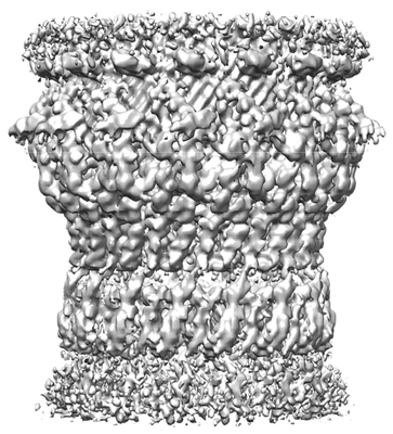 emd_6678.png emd_6678.png | 95.8 KB | ||
| Others | emd_6678_additional.map.gz | 165.6 MB | ||
| Archive directory |  http://ftp.pdbj.org/pub/emdb/structures/EMD-6678ftp://ftp.pdbj.org/pub/emdb/structures/EMD-6678 http://ftp.pdbj.org/pub/emdb/structures/EMD-6678ftp://ftp.pdbj.org/pub/emdb/structures/EMD-6678 | HTTPS FTP |
-Related structure data
| Related structure data |  6675C  6676C  6677C  5wq7C  5wq8C  5wq9C C: citing same article ( |
|---|---|
| Similar structure data |


-Links
| EMDB pages | EMDB (EBI/PDBe) / EMDataResource |
|---|---|
| Related items in Molecule of the Month |

-Map
| File | Download / File: emd_6678.map.gz / Format: CCP4 / Size: 209.3 MB / Type: IMAGE STORED AS FLOATING POINT NUMBER (4 BYTES) | ||||||||||||||||||||||||||||||||||||||||||||||||||||||||||||
|---|---|---|---|---|---|---|---|---|---|---|---|---|---|---|---|---|---|---|---|---|---|---|---|---|---|---|---|---|---|---|---|---|---|---|---|---|---|---|---|---|---|---|---|---|---|---|---|---|---|---|---|---|---|---|---|---|---|---|---|---|---|
| Annotation | VC_GspD_DelCap post-processing map | ||||||||||||||||||||||||||||||||||||||||||||||||||||||||||||
| Voxel size | X=Y=Z: 1.32 Å | ||||||||||||||||||||||||||||||||||||||||||||||||||||||||||||
| Density |
| ||||||||||||||||||||||||||||||||||||||||||||||||||||||||||||
| Symmetry | Space group: 1 | ||||||||||||||||||||||||||||||||||||||||||||||||||||||||||||
| Details | EMDB XML:
CCP4 map header:
| ||||||||||||||||||||||||||||||||||||||||||||||||||||||||||||
-Supplemental data
-Additional map: VC GspD DelCap unpost-processing map
| File | emd_6678_additional.map | ||||||||||||
|---|---|---|---|---|---|---|---|---|---|---|---|---|---|
| Annotation | VC_GspD_DelCap unpost-processing map | ||||||||||||
| Projections & Slices |
| ||||||||||||
| Density Histograms |
 Z
Z Y
Y X
X


- Sample components
-Entire : T2SS secretin GspD cap deletion
| Entire | Name: T2SS secretin GspD cap deletion |
|---|---|
| Components |
|
-Supramolecule #1: T2SS secretin GspD cap deletion
| Supramolecule | Name: T2SS secretin GspD cap deletion / type: complex / ID: 1 / Parent: 0 / Macromolecule list: all |
|---|---|
| Source (natural) | Organism: Vibrio cholerae O1 biovar El Tor str. N16961 (bacteria) |
-Supramolecule #2: T2SS secretin GspD cap deletion
| Supramolecule | Name: T2SS secretin GspD cap deletion / type: complex / ID: 2 / Parent: 1 / Macromolecule list: all |
|---|
-Macromolecule #1: T2SS secretin GspD cap deletion
| Macromolecule | Name: T2SS secretin GspD cap deletion / type: protein_or_peptide / ID: 1 / Enantiomer: LEVO |
|---|---|
| Sequence | String: MKYWLKKSSW LLAGSLLSTP LAMANEFSAS FKGTDIQEFI NIVGRNLEKT IIVDPSVRGK VDVRSFDTLN EEQYYSFFLS VLEVYGFAV VEMDNGVLKV IKSKDAKTSA IPVLSGEERA NGDEVITQVV AVKNVSVREL SPLLRQLIDN AGAGNVVHYD P ANIILITG ...String: MKYWLKKSSW LLAGSLLSTP LAMANEFSAS FKGTDIQEFI NIVGRNLEKT IIVDPSVRGK VDVRSFDTLN EEQYYSFFLS VLEVYGFAV VEMDNGVLKV IKSKDAKTSA IPVLSGEERA NGDEVITQVV AVKNVSVREL SPLLRQLIDN AGAGNVVHYD P ANIILITG RAAVVNRLAE IIRRVDQAGD KEIEVVELNN ASAAEMVRIV EALNKTTDAQ NTPEFLKPKF VADERTNSIL IS GDPKVRE RLKRLIKQLD VEMAAKGNNR VVYLKYAKAE DLVEVLKGVS ENLQAEKGTG QPTTSKRNEV MIAAHADTNS LVL TAPQDI MNAMLEVIGQ LDIRRAQVLI EALIVEMAEG DGINLGVQWG SLESGSVIQY GNTGASIGNV MIGLEEAKKG DYTK LASAL SSIQGAAVSI AMGDWTALIN AVSNDSSSNI LSSPSITVMD NGEASFIVGE EVPVITGSTA GSNNDNPFQT VDRKE VGIK LKVVPQINEG NSVQLNIEQE VSNVLGANGA VDVRFAKRQL NTSVMVQDGQ MLVLGGLIDE RALESESKVP LLGDIP LLG QLFRSTSSQV EKKNLMVFIK PTIIRDGVTA DGITQRKYNY IRAEQLFRAE KGLRLLDDAS VPVLPKFGDD RRHSPEI QA FIEQMEAKQ |
-Experimental details
-Structure determination
| Method | cryo EM |
|---|---|
 Processing Processing | single particle reconstruction |
| Aggregation state | particle |
-Sample preparation
| Buffer | pH: 8 |
|---|---|
| Vitrification | Cryogen name: ETHANE |
- Electron microscopy
| Microscope | FEI TITAN KRIOS |
|---|---|
| Electron beam | Acceleration voltage: 300 kV / Electron source: FIELD EMISSION GUN |
| Electron optics | Illumination mode: OTHER / Imaging mode: DARK FIELD |
| Image recording | Film or detector model: GATAN K2 SUMMIT (4k x 4k) / Average electron dose: 1.6 e/Å2 |
| Experimental equipment |  Model: Titan Krios / Image courtesy: FEI Company |
-Image processing
| Initial angle assignment | Type: RANDOM ASSIGNMENT |
|---|---|
| Final angle assignment | Type: OTHER |
| Final reconstruction | Applied symmetry - Point group: C15 (15 fold cyclic) / Resolution.type: BY AUTHOR / Resolution: 4.18 Å / Resolution method: FSC 0.143 CUT-OFF / Software - Name: Relion / Number images used: 3957 |
