 Movie
Movie Controller

[English] 日本語
 Yorodumi
- EMDB-6492: Electron cryo-microscopy of bacteriophage EL chaperonin in the AT... -
+ Open data

- Basic information
| Entry | Database: EMDB / ID: EMD-6492 | |||||||||
|---|---|---|---|---|---|---|---|---|---|---|
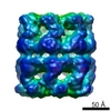
| Title | Electron cryo-microscopy of bacteriophage EL chaperonin in the ATP-bound conformation | |||||||||
 Map data Map data | Phi-EL chaperonin in the ATP-bound conformation | |||||||||
 Sample Sample |
| |||||||||
 Keywords Keywords |  chaperonin / phi-EL / protein folding / ATP conformation chaperonin / phi-EL / protein folding / ATP conformation | |||||||||
| Function / homology |  Function and homology information Function and homology informationATP-dependent protein folding chaperone / protein refolding / ATP binding / identical protein bindingSimilarity search - Function | |||||||||
| Biological species |  Pseudomonas phage EL (virus) Pseudomonas phage EL (virus) | |||||||||
| Method | single particle reconstruction / cryo EM / Resolution: 6.8 Å | |||||||||
 Authors Authors | Molugu SK / Hildenbrand ZL / Morgan DG / Sherman MB / He L / Georgopoulos C / Sernova NV / Kurochkina LP / Mesyanzhinov VV / Miroshnikov KA / Bernal RA | |||||||||
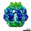
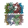
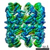
 Citation Citation | Journal: Structure / Year: 2016 Title: Ring Separation Highlights the Protein-Folding Mechanism Used by the Phage EL-Encoded Chaperonin. Authors: Sudheer K Molugu / Zacariah L Hildenbrand / David Gene Morgan / Michael B Sherman / Lilin He / Costa Georgopoulos / Natalia V Sernova / Lidia P Kurochkina / Vadim V Mesyanzhinov / Konstantin ...Authors: Sudheer K Molugu / Zacariah L Hildenbrand / David Gene Morgan / Michael B Sherman / Lilin He / Costa Georgopoulos / Natalia V Sernova / Lidia P Kurochkina / Vadim V Mesyanzhinov / Konstantin A Miroshnikov / Ricardo A Bernal /   Abstract: Chaperonins are ubiquitous, ATP-dependent protein-folding molecular machines that are essential for all forms of life. Bacteriophage φEL encodes its own chaperonin to presumably fold exceedingly ...Chaperonins are ubiquitous, ATP-dependent protein-folding molecular machines that are essential for all forms of life. Bacteriophage φEL encodes its own chaperonin to presumably fold exceedingly large viral proteins via profoundly different nucleotide-binding conformations. Our structural investigations indicate that ATP likely binds to both rings simultaneously and that a misfolded substrate acts as the trigger for ATP hydrolysis. More importantly, the φEL complex dissociates into two single rings resulting from an evolutionarily altered residue in the highly conserved ATP-binding pocket. Conformational changes also more than double the volume of the single-ring internal chamber such that larger viral proteins are accommodated. This is illustrated by the fact that φEL is capable of folding β-galactosidase, a 116-kDa protein. Collectively, the architecture and protein-folding mechanism of the φEL chaperonin are significantly different from those observed in group I and II chaperonins. | |||||||||
| History |
|
- Structure visualization
| Movie |
Movie viewer |
|---|---|
| Structure viewer | EM map: SurfViewMolmilJmol/JSmol |
| Supplemental images |

- Downloads & links
-EMDB archive
| Map data | emd_6492.map.gz | 7.4 MB | EMDB map data format | |
|---|---|---|---|---|
| Header (meta data) | emd-6492-v30.xmlemd-6492.xml | 10.4 KB 10.4 KB | Display Display | EMDB header |
| Images | 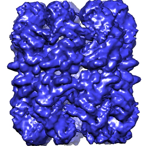 emd_6492.jpg emd_6492.jpg | 115.9 KB | ||
| Archive directory |  http://ftp.pdbj.org/pub/emdb/structures/EMD-6492ftp://ftp.pdbj.org/pub/emdb/structures/EMD-6492 http://ftp.pdbj.org/pub/emdb/structures/EMD-6492ftp://ftp.pdbj.org/pub/emdb/structures/EMD-6492 | HTTPS FTP |
-Related structure data


-Links
| EMDB pages | EMDB (EBI/PDBe) / EMDataResource |
|---|---|
| Related items in Molecule of the Month |

-Map
| File | Download / File: emd_6492.map.gz / Format: CCP4 / Size: 7.8 MB / Type: IMAGE STORED AS FLOATING POINT NUMBER (4 BYTES) | ||||||||||||||||||||||||||||||||||||||||||||||||||||||||||||
|---|---|---|---|---|---|---|---|---|---|---|---|---|---|---|---|---|---|---|---|---|---|---|---|---|---|---|---|---|---|---|---|---|---|---|---|---|---|---|---|---|---|---|---|---|---|---|---|---|---|---|---|---|---|---|---|---|---|---|---|---|---|
| Annotation | Phi-EL chaperonin in the ATP-bound conformation | ||||||||||||||||||||||||||||||||||||||||||||||||||||||||||||
| Voxel size | X=Y=Z: 2.4 Å | ||||||||||||||||||||||||||||||||||||||||||||||||||||||||||||
| Density |
| ||||||||||||||||||||||||||||||||||||||||||||||||||||||||||||
| Symmetry | Space group: 1 | ||||||||||||||||||||||||||||||||||||||||||||||||||||||||||||
| Details | EMDB XML:
CCP4 map header:
| ||||||||||||||||||||||||||||||||||||||||||||||||||||||||||||
-Supplemental data
- Sample components
-Entire : Bacteriphage phi-EL encoded chaperonin in the ATP-bound conformation
| Entire | Name: Bacteriphage phi-EL encoded chaperonin in the ATP-bound conformation |
|---|---|
| Components |
|
-Supramolecule #1000: Bacteriphage phi-EL encoded chaperonin in the ATP-bound conformation
| Supramolecule | Name: Bacteriphage phi-EL encoded chaperonin in the ATP-bound conformation type: sample / ID: 1000 / Oligomeric state: homotetradecamer / Number unique components: 1 |
|---|---|
| Molecular weight | Theoretical: 863.5354 KDa |
-Macromolecule #1: phi-EL chaperonin
| Macromolecule | Name: phi-EL chaperonin / type: protein_or_peptide / ID: 1 / Number of copies: 14 / Oligomeric state: tetradecamer / Recombinant expression: Yes |
|---|---|
| Source (natural) | Organism: Pseudomonas phage EL (virus) / synonym: Bacteriophage EL |
| Molecular weight | Theoretical: 61.6811 KDa |
| Recombinant expression | Organism:  Escherichia coli (E. coli) / Recombinant plasmid: pET22 Escherichia coli (E. coli) / Recombinant plasmid: pET22 |
| Sequence | UniProtKB: Putative GroEL-like chaperonine protein |
-Experimental details
-Structure determination
| Method | cryo EM |
|---|---|
 Processing Processing | single particle reconstruction |
| Aggregation state | particle |
-Sample preparation
| Concentration | 2.5 mg/mL |
|---|---|
| Buffer | pH: 7.5 Details: 50 mM HEPES, pH 7.5, 150 mM NaCl, 2 mM ATP, 2 mM MgCl2, 2 mM EDTA |
| Grid | Details: Quantifoil R2/2 grids glow-discharged in air for 1 minute |
| Vitrification | Cryogen name: ETHANE / Chamber humidity: 80 % / Chamber temperature: 100 K / Instrument: HOMEMADE PLUNGER / Method: Blot for 2-3 seconds before plunging |
- Electron microscopy
| Microscope | FEI TECNAI F20 |
|---|---|
| Electron beam | Acceleration voltage: 200 kV / Electron source: FIELD EMISSION GUN |
| Electron optics | Calibrated magnification: 50000 / Illumination mode: FLOOD BEAM / Imaging mode: BRIGHT FIELDBright-field microscopy / Nominal defocus max: 4.0 µm / Nominal defocus min: 1.5 µm |
| Sample stage | Specimen holder model: GATAN LIQUID NITROGEN |
| Temperature | Min: 88 K / Max: 103 K / Average: 100 K |
| Date | Aug 1, 2006 |
| Image recording | Category: FILM / Film or detector model: KODAK SO-163 FILM / Digitization - Scanner: OTHER / Digitization - Sampling interval: 12 µm / Number real images: 50 / Average electron dose: 10 e/Å2 / Bits/pixel: 8 |
| Experimental equipment |  Model: Tecnai F20 / Image courtesy: FEI Company |
-Image processing
| CTF correction | Details: Each particle |
|---|---|
| Final reconstruction | Algorithm: OTHER / Resolution.type: BY AUTHOR / Resolution: 6.8 Å / Resolution method: OTHER / Software - Name: EMAN, EMAN2, RELION / Number images used: 40613 |
