 Movie
Movie Controller

[English] 日本語
 Yorodumi
- EMDB-6337: Structure of the L-protein of vesicular stomatitis virus from ele... -
+ Open data

- Basic information
| Entry | Database: EMDB / ID: EMD-6337 | |||||||||
|---|---|---|---|---|---|---|---|---|---|---|
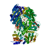
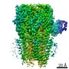
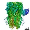
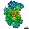
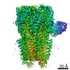
| Title | Structure of the L-protein of vesicular stomatitis virus from electron cryomicroscopy | |||||||||
 Map data Map data | Reconstruction of the L-protein of vesicular stomatitis virus | |||||||||
 Sample Sample |
| |||||||||
 Keywords Keywords |  RNA-dependent RNA polymerase / RNA capping / cryoEM single-particle analysis RNA-dependent RNA polymerase / RNA capping / cryoEM single-particle analysis | |||||||||
| Function / homology |  Function and homology information Function and homology informationNNS virus cap methyltransferase / GDP polyribonucleotidyltransferase / negative stranded viral RNA replication / viral transcription / Hydrolases; Acting on acid anhydrides; In phosphorus-containing anhydrides / virion component / mRNA 5'-cap (guanine-N7-)-methyltransferase activity / host cell cytoplasm / RNA-directed RNA polymerase / RNA-dependent RNA polymerase activity ...NNS virus cap methyltransferase / GDP polyribonucleotidyltransferase / negative stranded viral RNA replication / viral transcription / Hydrolases; Acting on acid anhydrides; In phosphorus-containing anhydrides / virion component / mRNA 5'-cap (guanine-N7-)-methyltransferase activity / host cell cytoplasm / RNA-directed RNA polymerase / RNA-dependent RNA polymerase activity / GTPase activity / ATP binding / metal ion bindingSimilarity search - Function | |||||||||
| Biological species |  Vesicular stomatitis virus Vesicular stomatitis virus | |||||||||
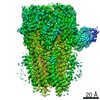
| Method | single particle reconstruction / cryo EM / Resolution: 3.8 Å | |||||||||
 Authors Authors | Liang B / Li Z / Jenni S / Rameh AA / Morin BM / Grant T / Grigorieff N / Harrison SC / Whelan SPJ | |||||||||
 Citation Citation | Journal: Cell / Year: 2015 Title: Structure of the L Protein of Vesicular Stomatitis Virus from Electron Cryomicroscopy. Authors: Bo Liang / Zongli Li / Simon Jenni / Amal A Rahmeh / Benjamin M Morin / Timothy Grant / Nikolaus Grigorieff / Stephen C Harrison / Sean P J Whelan /  Abstract: The large (L) proteins of non-segmented, negative-strand RNA viruses, a group that includes Ebola and rabies viruses, catalyze RNA-dependent RNA polymerization with viral ribonucleoprotein as ...The large (L) proteins of non-segmented, negative-strand RNA viruses, a group that includes Ebola and rabies viruses, catalyze RNA-dependent RNA polymerization with viral ribonucleoprotein as template, a non-canonical sequence of capping and methylation reactions, and polyadenylation of viral messages. We have determined by electron cryomicroscopy the structure of the vesicular stomatitis virus (VSV) L protein. The density map, at a resolution of 3.8 Å, has led to an atomic model for nearly all of the 2109-residue polypeptide chain, which comprises three enzymatic domains (RNA-dependent RNA polymerase [RdRp], polyribonucleotidyl transferase [PRNTase], and methyltransferase) and two structural domains. The RdRp resembles the corresponding enzymatic regions of dsRNA virus polymerases and influenza virus polymerase. A loop from the PRNTase (capping) domain projects into the catalytic site of the RdRp, where it appears to have the role of a priming loop and to couple product elongation to large-scale conformational changes in L. | |||||||||
| History |
|
- Structure visualization
| Movie |
Movie viewer |
|---|---|
| Structure viewer | EM map: SurfViewMolmilJmol/JSmol |
| Supplemental images |

- Downloads & links
-EMDB archive
| Map data | emd_6337.map.gz | 6.2 MB | EMDB map data format | |
|---|---|---|---|---|
| Header (meta data) | emd-6337-v30.xmlemd-6337.xml | 11.3 KB 11.3 KB | Display Display | EMDB header |
| Images | 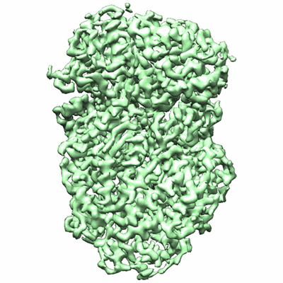 400_6337.gif 400_6337.gif 80_6337.gif 80_6337.gif | 82.4 KB 5 KB | ||
| Filedesc structureFactors | emd_6337_sf.cif.gz | 1.2 MB | ||
| Archive directory |  http://ftp.pdbj.org/pub/emdb/structures/EMD-6337ftp://ftp.pdbj.org/pub/emdb/structures/EMD-6337 http://ftp.pdbj.org/pub/emdb/structures/EMD-6337ftp://ftp.pdbj.org/pub/emdb/structures/EMD-6337 | HTTPS FTP |
-Related structure data
| Related structure data | 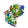 5a22MC M: atomic model generated by this map C: citing same article ( |
|---|---|
| Similar structure data |
-Links
| EMDB pages | EMDB (EBI/PDBe) / EMDataResource |
|---|---|
| Related items in Molecule of the Month |

-Map
| File | Download / File: emd_6337.map.gz / Format: CCP4 / Size: 6.4 MB / Type: IMAGE STORED AS FLOATING POINT NUMBER (4 BYTES) | ||||||||||||||||||||||||||||||||||||||||||||||||||||||||||||
|---|---|---|---|---|---|---|---|---|---|---|---|---|---|---|---|---|---|---|---|---|---|---|---|---|---|---|---|---|---|---|---|---|---|---|---|---|---|---|---|---|---|---|---|---|---|---|---|---|---|---|---|---|---|---|---|---|---|---|---|---|---|
| Annotation | Reconstruction of the L-protein of vesicular stomatitis virus | ||||||||||||||||||||||||||||||||||||||||||||||||||||||||||||
| Voxel size | X=Y=Z: 1.237 Å | ||||||||||||||||||||||||||||||||||||||||||||||||||||||||||||
| Density |
| ||||||||||||||||||||||||||||||||||||||||||||||||||||||||||||
| Symmetry | Space group: 1 | ||||||||||||||||||||||||||||||||||||||||||||||||||||||||||||
| Details | EMDB XML:
CCP4 map header:
| ||||||||||||||||||||||||||||||||||||||||||||||||||||||||||||
-Supplemental data
- Sample components
-Entire : VSV-L
| Entire | Name: VSV-L |
|---|---|
| Components |
|
-Supramolecule #1000: VSV-L
| Supramolecule | Name: VSV-L / type: sample / ID: 1000 Oligomeric state: One monomer of VSV-L bound to one monomer of P Number unique components: 2 |
|---|---|
| Molecular weight | Theoretical: 250 KDa |
-Macromolecule #1: VSV large protein
| Macromolecule | Name: VSV large protein / type: protein_or_peptide / ID: 1 / Name.synonym: VSV-L / Number of copies: 1 / Oligomeric state: Monomer / Recombinant expression: Yes |
|---|---|
| Source (natural) | Organism: Vesicular stomatitis virus |
| Molecular weight | Theoretical: 240 KDa |
| Recombinant expression | Organism:   Spodoptera frugiperda (fall armyworm) / Recombinant cell: Sf21 Spodoptera frugiperda (fall armyworm) / Recombinant cell: Sf21 |
-Macromolecule #2: VSV phosphoprotein
| Macromolecule | Name: VSV phosphoprotein / type: protein_or_peptide / ID: 2 / Name.synonym: P / Number of copies: 1 / Oligomeric state: Monomer / Recombinant expression: Yes |
|---|---|
| Source (natural) | Organism: Vesicular stomatitis virus |
| Molecular weight | Theoretical: 10 KDa |
| Recombinant expression | Organism:  Escherichia coli BL21 (bacteria) / Recombinant strain: BL21 Escherichia coli BL21 (bacteria) / Recombinant strain: BL21 |
-Experimental details
-Structure determination
| Method | cryo EM |
|---|---|
 Processing Processing | single particle reconstruction |
| Aggregation state | particle |
-Sample preparation
| Concentration | 0.35 mg/mL |
|---|---|
| Buffer | pH: 7.4 / Details: 25 mM HEPES, 250 mM NaCl, 6 mM MgSO4, 0.5 mM TCEP |
| Grid | Details: 400 mesh Quantifoil R1.2/1.3 Cu grid |
| Vitrification | Cryogen name: ETHANE / Chamber humidity: 65 % / Instrument: FEI VITROBOT MARK I Method: Blot time 2 seconds, drain time 1 second before plunging |
- Electron microscopy
| Microscope | FEI TECNAI F20 |
|---|---|
| Electron beam | Acceleration voltage: 200 kV / Electron source: FIELD EMISSION GUN |
| Electron optics | Calibrated magnification: 40410 / Illumination mode: FLOOD BEAM / Imaging mode: BRIGHT FIELDBright-field microscopy / Cs: 2.0 mm / Nominal defocus max: 2.3 µm / Nominal defocus min: 0.9 µm / Nominal magnification: 29000 |
| Sample stage | Specimen holder: CT3500 / Specimen holder model: GATAN LIQUID NITROGEN |
| Details | Beam intensity: 8 electrons/pixel/s Movie mode: 30 frames, 5 frames/s |
| Date | Aug 1, 2014 |
| Image recording | Category: CCD / Film or detector model: GATAN K2 (4k x 4k) / Digitization - Sampling interval: 5 µm / Number real images: 1272 / Average electron dose: 31 e/Å2 Details: Images are the sums of all 30 aligned movie frames (high dose) or frames 3 - 12 (low-dose). |
| Experimental equipment |  Model: Tecnai F20 / Image courtesy: FEI Company |
-Image processing
| CTF correction | Details: Each particle |
|---|---|
| Final two d classification | Number classes: 3 |
| Final reconstruction | Algorithm: OTHER / Resolution.type: BY AUTHOR / Resolution: 3.8 Å / Resolution method: OTHER / Software - Name: EMAN2, IMAGIC, Frealign Details: The final map represents the best class out of three classes. Number images used: 74940 |
| Details | An initial map was obtained with EMAN2, IMAGIC, and TIGRIS. CTF was determined using CTFFIND3. Refinement and classification were done using Frealign. |
