Movie
Movie Controller


+ Open data

- Basic information
| Entry | Database: EMDB / ID: EMD-5679 | |||||||||
|---|---|---|---|---|---|---|---|---|---|---|
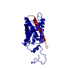
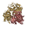
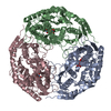
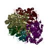
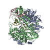
| Title | Electron Microscopy of the Aquaporin-0/Calmodulin Complex | |||||||||
 Map data Map data | 3D Reconstruction of the Aquaporin-0/Calmodulin Complex | |||||||||
 Sample Sample |
| |||||||||
 Keywords Keywords | aquaporin / calmodulin / calcium regulation / water channel / membrane protein complex / electron microscopy | |||||||||
| Function / homology |  Function and homology information Function and homology informationgap junction-mediated intercellular transport / water transport / water channel activity / : / structural constituent of eye lens / establishment of protein localization to mitochondrial membrane / gap junction / type 3 metabotropic glutamate receptor binding / CaM pathway / Cam-PDE 1 activation ...gap junction-mediated intercellular transport / water transport / water channel activity / : / structural constituent of eye lens / establishment of protein localization to mitochondrial membrane / gap junction / type 3 metabotropic glutamate receptor binding / CaM pathway / Cam-PDE 1 activation / Sodium/Calcium exchangers / lens development in camera-type eye / Calmodulin induced events / regulation of synaptic vesicle endocytosis / Reduction of cytosolic Ca++ levels / CREB1 phosphorylation through the activation of CaMKII/CaMKK/CaMKIV cascasde / Activation of Ca-permeable Kainate Receptor / Loss of phosphorylation of MECP2 at T308 / CREB1 phosphorylation through the activation of Adenylate Cyclase / PKA activation / negative regulation of high voltage-gated calcium channel activity / CaMK IV-mediated phosphorylation of CREB / Glycogen breakdown (glycogenolysis) / positive regulation of cyclic-nucleotide phosphodiesterase activity / regulation of synaptic vesicle exocytosis / organelle localization by membrane tethering / negative regulation of calcium ion export across plasma membrane / CLEC7A (Dectin-1) induces NFAT activation / mitochondrion-endoplasmic reticulum membrane tethering / autophagosome membrane docking / regulation of cardiac muscle cell action potential / Activation of RAC1 downstream of NMDARs / response to corticosterone / positive regulation of DNA binding / positive regulation of ryanodine-sensitive calcium-release channel activity / nitric-oxide synthase binding / regulation of cell communication by electrical coupling involved in cardiac conduction / Negative regulation of NMDA receptor-mediated neuronal transmission / negative regulation of peptidyl-threonine phosphorylation / Synthesis of IP3 and IP4 in the cytosol / Unblocking of NMDA receptors, glutamate binding and activation / Phase 0 - rapid depolarisation / negative regulation of ryanodine-sensitive calcium-release channel activity / protein phosphatase activator activity / RHO GTPases activate PAKs / : / Ion transport by P-type ATPases / Long-term potentiation / Uptake and function of anthrax toxins / Regulation of MECP2 expression and activity / Calcineurin activates NFAT / adenylate cyclase binding / catalytic complex / positive regulation of cell adhesion / DARPP-32 events / detection of calcium ion / regulation of cardiac muscle contraction / regulation of ryanodine-sensitive calcium-release channel activity / Smooth Muscle Contraction / cellular response to interferon-beta / RHO GTPases activate IQGAPs / regulation of cardiac muscle contraction by regulation of the release of sequestered calcium ion / calcium channel inhibitor activity / Protein methylation / phosphatidylinositol 3-kinase binding / eNOS activation / enzyme regulator activity / activation of adenylate cyclase activity / regulation of release of sequestered calcium ion into cytosol by sarcoplasmic reticulum / Activation of AMPK downstream of NMDARs / Tetrahydrobiopterin (BH4) synthesis, recycling, salvage and regulation / : / Ion homeostasis / titin binding / regulation of calcium-mediated signaling / positive regulation of protein autophosphorylation / voltage-gated potassium channel complex / sperm midpiece / calcium channel complex / response to amphetamine / substantia nigra development / visual perception / adenylate cyclase activator activity / Ras activation upon Ca2+ influx through NMDA receptor / nitric-oxide synthase regulator activity / regulation of heart rate / sarcomere / FCERI mediated Ca+2 mobilization / protein serine/threonine kinase activator activity / FCGR3A-mediated IL10 synthesis / VEGFR2 mediated vascular permeability / Antigen activates B Cell Receptor (BCR) leading to generation of second messengers / VEGFR2 mediated cell proliferation / regulation of cytokinesis / positive regulation of peptidyl-threonine phosphorylation / positive regulation of nitric-oxide synthase activity / Translocation of SLC2A4 (GLUT4) to the plasma membrane / spindle microtubule / RAF activation / positive regulation of receptor signaling pathway via JAK-STAT Similarity search - Function | |||||||||
| Biological species |   Homo sapiens (human) Homo sapiens (human) | |||||||||
| Method | single particle reconstruction / negative staining / Resolution: 25.0 Å | |||||||||
 Authors Authors | Reichow SL / Clemens DM / Freites JA / Nemeth-Cahalan KL / Heyden M / Tobias DJ / Hall JE / Gonen T | |||||||||
 Citation Citation | Journal: Nat Struct Mol Biol / Year: 2013 Title: Allosteric mechanism of water-channel gating by Ca2+-calmodulin. Authors: Steve L Reichow / Daniel M Clemens / J Alfredo Freites / Karin L Németh-Cahalan / Matthias Heyden / Douglas J Tobias / James E Hall / Tamir Gonen /  Abstract: Calmodulin (CaM) is a universal regulatory protein that communicates the presence of calcium to its molecular targets and correspondingly modulates their function. This key signaling protein is ...Calmodulin (CaM) is a universal regulatory protein that communicates the presence of calcium to its molecular targets and correspondingly modulates their function. This key signaling protein is important for controlling the activity of hundreds of membrane channels and transporters. However, understanding of the structural mechanisms driving CaM regulation of full-length membrane proteins has remained elusive. In this study, we determined the pseudoatomic structure of full-length mammalian aquaporin-0 (AQP0, Bos taurus) in complex with CaM, using EM to elucidate how this signaling protein modulates water-channel function. Molecular dynamics and functional mutation studies reveal how CaM binding inhibits AQP0 water permeability by allosterically closing the cytoplasmic gate of AQP0. Our mechanistic model provides new insight, only possible in the context of the fully assembled channel, into how CaM regulates multimeric channels by facilitating cooperativity between adjacent subunits. | |||||||||
| History |
|
- Structure visualization
| Movie |
Movie viewer |
|---|---|
| Structure viewer | EM map: SurfViewMolmilJmol/JSmol |
| Supplemental images |
 UCSF Chimera
- Downloads & links
-EMDB archive
| Map data | emd_5679.map.gz | 458.7 KB | EMDB map data format | |
|---|---|---|---|---|
| Header (meta data) | emd-5679-v30.xmlemd-5679.xml | 13.2 KB 13.2 KB | Display Display | EMDB header |
| Images | emd_5679.tif | 287.7 KB | ||
| Archive directory |  http://ftp.pdbj.org/pub/emdb/structures/EMD-5679ftp://ftp.pdbj.org/pub/emdb/structures/EMD-5679 http://ftp.pdbj.org/pub/emdb/structures/EMD-5679ftp://ftp.pdbj.org/pub/emdb/structures/EMD-5679 | HTTPS FTP |
-Validation report
| Summary document | emd_5679_validation.pdf.gz | 285.4 KB | Display | EMDB validaton report |
|---|---|---|---|---|
| Full document | emd_5679_full_validation.pdf.gz | 285 KB | Display | |
| Data in XML | emd_5679_validation.xml.gz | 5.1 KB | Display | |
| Arichive directory | https://ftp.pdbj.org/pub/emdb/validation_reports/EMD-5679ftp://ftp.pdbj.org/pub/emdb/validation_reports/EMD-5679 | HTTPS FTP |
-Related structure data
| Related structure data |  3j41MC M: atomic model generated by this map C: citing same article ( |
|---|---|
| Similar structure data |


-Links
| EMDB pages | EMDB (EBI/PDBe) / EMDataResource |
|---|---|
| Related items in Molecule of the Month |


-Map
| File | Download / File: emd_5679.map.gz / Format: CCP4 / Size: 1.1 MB / Type: IMAGE STORED AS FLOATING POINT NUMBER (4 BYTES) | ||||||||||||||||||||||||||||||||||||||||||||||||||||||||||||||||||||
|---|---|---|---|---|---|---|---|---|---|---|---|---|---|---|---|---|---|---|---|---|---|---|---|---|---|---|---|---|---|---|---|---|---|---|---|---|---|---|---|---|---|---|---|---|---|---|---|---|---|---|---|---|---|---|---|---|---|---|---|---|---|---|---|---|---|---|---|---|---|
| Annotation | 3D Reconstruction of the Aquaporin-0/Calmodulin Complex | ||||||||||||||||||||||||||||||||||||||||||||||||||||||||||||||||||||
| Voxel size | X=Y=Z: 3.98 Å | ||||||||||||||||||||||||||||||||||||||||||||||||||||||||||||||||||||
| Density |
| ||||||||||||||||||||||||||||||||||||||||||||||||||||||||||||||||||||
| Symmetry | Space group: 1 | ||||||||||||||||||||||||||||||||||||||||||||||||||||||||||||||||||||
| Details | EMDB XML:
CCP4 map header:
| ||||||||||||||||||||||||||||||||||||||||||||||||||||||||||||||||||||
-Supplemental data
- Sample components
-Entire : Aquaporin-0 bound to Calmodulin
| Entire | Name: Aquaporin-0 bound to Calmodulin |
|---|---|
| Components |
|
-Supramolecule #1000: Aquaporin-0 bound to Calmodulin
| Supramolecule | Name: Aquaporin-0 bound to Calmodulin / type: sample / ID: 1000 Details: Sample was prepared for electron microscopy with negative stain Oligomeric state: One tetramer of Aquaporin-0 bound to 2 molecules of Calmodulin Number unique components: 2 |
|---|---|
| Molecular weight | Experimental: 130 KDa / Theoretical: 130 KDa / Method: Size-exclusion Chromatography and SDS-PAGE |
-Macromolecule #1: Aquaporin-0
| Macromolecule | Name: Aquaporin-0 / type: protein_or_peptide / ID: 1 / Name.synonym: AQP0, MIP / Details: Crosslinked to Calmodulin using EDC/NHS / Number of copies: 4 / Oligomeric state: tetramer / Recombinant expression: No / Database: NCBI |
|---|---|
| Source (natural) | Organism: |
| Molecular weight | Experimental: 25 KDa / Theoretical: 25 KDa |
| Sequence | UniProtKB: Pas12 / InterPro: Major intrinsic protein |
-Macromolecule #2: Calmodulin
| Macromolecule | Name: Calmodulin / type: protein_or_peptide / ID: 2 / Name.synonym: CaM / Details: Calmodulin crosslinked to Aquaporin-0 / Number of copies: 2 / Oligomeric state: monomer / Recombinant expression: Yes |
|---|---|
| Source (natural) | Organism: Homo sapiens (human) / synonym: Human / Location in cell: cytoplasmic |
| Molecular weight | Experimental: 17 KDa / Theoretical: 17 KDa |
| Recombinant expression | Organism:  |
| Sequence | UniProtKB: Calmodulin-3 |
-Experimental details
-Structure determination
| Method | negative staining |
|---|---|
 Processing Processing | single particle reconstruction |
| Aggregation state | particle |
-Sample preparation
| Concentration | 0.02 mg/mL |
|---|---|
| Buffer | pH: 7.4 / Details: 25mM HEPES, 5mM CaCl2, 0.3% decylmaltoside |
| Staining | Type: NEGATIVE / Details: 0.75% uranyl formate |
| Grid | Details: 400 mesh carbon coated grid (Ted Pella) |
| Vitrification | Cryogen name: NONE / Instrument: OTHER |
- Electron microscopy
| Microscope | FEI TECNAI 12 |
|---|---|
| Alignment procedure | Legacy - Astigmatism: Objective lens astigmatism was corrected at 100,000 times magnification Legacy - Electron beam tilt params: 0 |
| Date | Feb 25, 2010 |
| Image recording | Category: FILM / Film or detector model: KODAK SO-163 FILM / Digitization - Scanner: NIKON SUPER COOLSCAN 9000 / Digitization - Sampling interval: 6.35 µm / Number real images: 200 / Average electron dose: 15 e/Å2 / Bits/pixel: 16 |
| Tilt angle min | 0 |
| Electron beam | Acceleration voltage: 120 kV / Electron source: LAB6 |
| Electron optics | Calibrated magnification: 52000 / Illumination mode: FLOOD BEAM / Imaging mode: BRIGHT FIELD / Cs: 2 mm / Nominal defocus max: 2.0 µm / Nominal defocus min: 1.0 µm / Nominal magnification: 52000 |
| Sample stage | Specimen holder model: OTHER / Tilt angle max: 50 |
-Image processing
| Details | Particles were selected from a tilted pair dataset at 0 and 50 degree tilt using SPIDER. An initial reconstruction was generated using random conical tilt methods in SPIDER and refined in FREALIGN |
|---|---|
| CTF correction | Details: CTF-TILT, each micrograph |
| Final reconstruction | Algorithm: OTHER / Resolution.type: BY AUTHOR / Resolution: 25.0 Å / Resolution method: FSC 0.5 CUT-OFF / Software - Name: SPIDER, FREALIGN Details: Final Map with C2 Symmetry and Filtered to 25 Angstrom Number images used: 11720 |