- EMDB-5666: Cryo-EM structure of beta-hydroxyhexaketide-PikAIII conformation 2 -
+
Open data
ID or keywords:
Loading...
-
Basic information
Entry
Database: EMDB / ID: EMD-5666
Title
Cryo-EM structure of beta-hydroxyhexaketide-PikAIII conformation 2
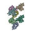
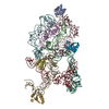
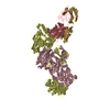
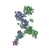
Map data
Reconstruction of the 5th module from the pikromycin biosynthetic pathway (PikAIII) incubated with NADPH, methylmalonyl-CoA, and thiophenol-pentaketide.
Sample
Sample: The 5th module from the pikromycin biosynthetic pathway (PikAIII) incubated with NADPH, methylmalonyl-CoA, and thiophenol-pentaketide
10-deoxymethynolide synthase / narbonolide synthase / macrolide biosynthetic process / acyltransferase activity, transferring groups other than amino-acyl groups / phosphopantetheine binding / 3-oxoacyl-[acyl-carrier-protein] synthase activity / fatty acid biosynthetic process / identical protein binding Similarity search - Function
Journal: Nature / Year: 2014 Title: Structural rearrangements of a polyketide synthase module during its catalytic cycle. Authors: Jonathan R Whicher / Somnath Dutta / Douglas A Hansen / Wendi A Hale / Joseph A Chemler / Annie M Dosey / Alison R H Narayan / Kristina Håkansson / David H Sherman / Janet L Smith / Georgios Skiniotis / Abstract: The polyketide synthase (PKS) mega-enzyme assembly line uses a modular architecture to synthesize diverse and bioactive natural products that often constitute the core structures or complete chemical ...The polyketide synthase (PKS) mega-enzyme assembly line uses a modular architecture to synthesize diverse and bioactive natural products that often constitute the core structures or complete chemical entities for many clinically approved therapeutic agents. The architecture of a full-length PKS module from the pikromycin pathway of Streptomyces venezuelae creates a reaction chamber for the intramodule acyl carrier protein (ACP) domain that carries building blocks and intermediates between acyltransferase, ketosynthase and ketoreductase active sites (see accompanying paper). Here we determine electron cryo-microscopy structures of a full-length pikromycin PKS module in three key biochemical states of its catalytic cycle. Each biochemical state was confirmed by bottom-up liquid chromatography/Fourier transform ion cyclotron resonance mass spectrometry. The ACP domain is differentially and precisely positioned after polyketide chain substrate loading on the active site of the ketosynthase, after extension to the β-keto intermediate, and after β-hydroxy product generation. The structures reveal the ACP dynamics for sequential interactions with catalytic domains within the reaction chamber, and for transferring the elongated and processed polyketide substrate to the next module in the PKS pathway. During the enzymatic cycle the ketoreductase domain undergoes dramatic conformational rearrangements that enable optimal positioning for reductive processing of the ACP-bound polyketide chain elongation intermediate. These findings have crucial implications for the design of functional PKS modules, and for the engineering of pathways to generate pharmacologically relevant molecules.
History
Deposition
May 2, 2013
-
Header (metadata) release
Jul 3, 2013
-
Map release
Jun 25, 2014
-
Update
Oct 22, 2014
-
Current status
Oct 22, 2014
Processing site: RCSB / Status: Released
-
Structure visualization
Movie
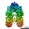
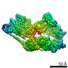
Surface view with section colored by density value
Download / File: emd_5666.map.gz / Format: CCP4 / Size: 26.4 MB / Type: IMAGE STORED AS FLOATING POINT NUMBER (4 BYTES)
Annotation
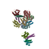
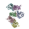
Reconstruction of the 5th module from the pikromycin biosynthetic pathway (PikAIII) incubated with NADPH, methylmalonyl-CoA, and thiophenol-pentaketide.
Voxel size
X=Y=Z: 2.24 Å
Density
Contour Level
By AUTHOR: 6.8 / Movie #1: 6.8
Minimum - Maximum
-10.71022892 - 31.39071083
Average (Standard dev.)
0.0 (±1.0)
Symmetry
Space group: 1
Details
EMDB XML:
Map geometry
Axis order
X
Y
Z
Origin
-16
-16
-16
Dimensions
192
192
192
Spacing
192
192
192
Cell
A=B=C: 430.08002 Å α=β=γ: 90.0 °
CCP4 map header:
mode
Image stored as Reals
Å/pix. X/Y/Z
2.24
2.24
2.24
M x/y/z
192
192
192
origin x/y/z
0.000
0.000
0.000
length x/y/z
430.080
430.080
430.080
α/β/γ
90.000
90.000
90.000
start NX/NY/NZ
-132
-122
-147
NX/NY/NZ
250
274
261
MAP C/R/S
1
2
3
start NC/NR/NS
-16
-16
-16
NC/NR/NS
192
192
192
D min/max/mean
-10.710
31.391
0.000
-
Supplemental data
-
Sample components
-
Entire : The 5th module from the pikromycin biosynthetic pathway (PikAIII)...
Entire
Name: The 5th module from the pikromycin biosynthetic pathway (PikAIII) incubated with NADPH, methylmalonyl-CoA, and thiophenol-pentaketide
Components
Sample: The 5th module from the pikromycin biosynthetic pathway (PikAIII) incubated with NADPH, methylmalonyl-CoA, and thiophenol-pentaketide
Supramolecule #1000: The 5th module from the pikromycin biosynthetic pathway (PikAIII)...
Supramolecule
Name: The 5th module from the pikromycin biosynthetic pathway (PikAIII) incubated with NADPH, methylmalonyl-CoA, and thiophenol-pentaketide type: sample / ID: 1000 Details: Sample was not frozen prior to loading on the grid. The sample was monodisperse. Oligomeric state: Dimer / Number unique components: 4
Cryogen name: ETHANE / Chamber humidity: 100 % / Chamber temperature: 89 K / Instrument: FEI VITROBOT MARK IV / Method: Blot for 1.5-2.0 seconds before plunging.
-
Electron microscopy
Microscope
FEI TECNAI F20
Electron beam
Acceleration voltage: 120 kV / Electron source: FIELD EMISSION GUN
In the structure databanks used in Yorodumi, some data are registered as the other names, "COVID-19 virus" and "2019-nCoV". Here are the details of the virus and the list of structure data.
Jan 31, 2019. EMDB accession codes are about to change! (news from PDBe EMDB page)
EMDB accession codes are about to change! (news from PDBe EMDB page)
The allocation of 4 digits for EMDB accession codes will soon come to an end. Whilst these codes will remain in use, new EMDB accession codes will include an additional digit and will expand incrementally as the available range of codes is exhausted. The current 4-digit format prefixed with “EMD-” (i.e. EMD-XXXX) will advance to a 5-digit format (i.e. EMD-XXXXX), and so on. It is currently estimated that the 4-digit codes will be depleted around Spring 2019, at which point the 5-digit format will come into force.
The EM Navigator/Yorodumi systems omit the EMD- prefix.
Related info.:Q: What is EMD? / ID/Accession-code notation in Yorodumi/EM Navigator
Yorodumi is a browser for structure data from EMDB, PDB, SASBDB, etc.
This page is also the successor to EM Navigator detail page, and also detail information page/front-end page for Omokage search.
The word "yorodu" (or yorozu) is an old Japanese word meaning "ten thousand". "mi" (miru) is to see.
Related info.:EMDB / PDB / SASBDB / Comparison of 3 databanks / Yorodumi Search / Aug 31, 2016. New EM Navigator & Yorodumi / Yorodumi Papers / Jmol/JSmol / Function and homology information / Changes in new EM Navigator and Yorodumi
 Movie
Movie Controller

 Yorodumi
 Open data
 Basic information
 Map data
 Sample
 Keywords
 Function and homology information
 phosphopantetheine binding / 3-oxoacyl-[acyl-carrier-protein] synthase activity / fatty acid biosynthetic process / identical protein binding
 Authors
 Citation
 Structure visualization
 Downloads & links
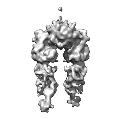 400_5666.gif
 80_5666.gif
 http://ftp.pdbj.org/pub/emdb/structures/EMD-5666


 Sample components
 Processing
 Electron microscopy