Movie
Movie Controller

[English] 日本語
 Yorodumi
- EMDB-5415: Structure of AAV-DJ, a Retargeted Gene Therapy Vector: Cryo-Elect... -
+ Open data

- Basic information
| Entry | Database: EMDB / ID: EMD-5415 | |||||||||
|---|---|---|---|---|---|---|---|---|---|---|
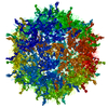
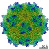
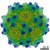
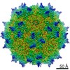
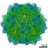
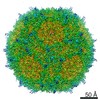
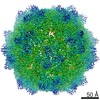
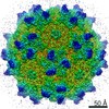
| Title | Structure of AAV-DJ, a Retargeted Gene Therapy Vector: Cryo-Electron Microscopy at 4.5A resolution | |||||||||
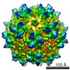
 Map data Map data | Reconstruction of AAV-DJ, a re-targeted gene therapy vector | |||||||||
 Sample Sample |
| |||||||||
 Keywords Keywords |  Gene therapy / adeno-associated virus / vector / cryo-electron microscopy / directed evolution / AAV Gene therapy / adeno-associated virus / vector / cryo-electron microscopy / directed evolution / AAV | |||||||||
| Function / homology | Phospholipase A2-like domain / Phospholipase A2-like domain / Parvovirus coat protein VP2 / Parvovirus coat protein VP1/VP2 / Parvovirus coat protein VP2 / Capsid/spike protein, ssDNA virus / T=1 icosahedral viral capsid / structural molecule activity / Capsid protein VP1 Function and homology information Function and homology information | |||||||||
| Biological species |   Adeno-associated virus Adeno-associated virus | |||||||||
| Method | single particle reconstruction / cryo EM / Resolution: 4.5 Å | |||||||||
 Authors Authors | Lerch TF / O'Donnell JK / Meyer NL / Xie Q Taylor KA / Stagg SC / Chapman MS | |||||||||
 Citation Citation | Journal: Structure / Year: 2012 Title: Structure of AAV-DJ, a retargeted gene therapy vector: cryo-electron microscopy at 4.5 Å resolution. Authors: Thomas F Lerch / Jason K O'Donnell / Nancy L Meyer / Qing Xie / Kenneth A Taylor / Scott M Stagg / Michael S Chapman /  Abstract: AAV-DJ, a leading candidate vector for liver gene therapy, was created through random homologous recombination followed by directed evolution, selecting for in vivo liver tropism and resistance to ...AAV-DJ, a leading candidate vector for liver gene therapy, was created through random homologous recombination followed by directed evolution, selecting for in vivo liver tropism and resistance to in vitro immune neutralization. Here, the 4.5 Å resolution cryo-EM structure is determined for the engineered AAV vector, revealing structural features that illuminate its phenotype. The heparan sulfate receptor-binding site is little changed from AAV-2, and heparin-binding affinity is similar. A loop that is antigenic in other serotypes has a unique conformation in AAV-DJ that would conflict with the binding of an AAV-2 neutralizing monoclonal antibody. This is consistent with increased resistance to neutralization by human polyclonal sera, raising the possibility that changed tropism may be a secondary effect of altered immune interactions. The reconstruction exemplifies analysis of fine structural changes and the potential of cryo-EM, in favorable cases, to characterize mutant or ligand-bound complexes. | |||||||||
| History |
|
- Structure visualization
| Movie |
Movie viewer |
|---|---|
| Structure viewer | EM map: SurfViewMolmilJmol/JSmol |
| Supplemental images |

- Downloads & links
-EMDB archive
| Map data | emd_5415.map.gz | 148.1 MB | EMDB map data format | |
|---|---|---|---|---|
| Header (meta data) | emd-5415-v30.xmlemd-5415.xml | 9.8 KB 9.8 KB | Display Display | EMDB header |
| Images | 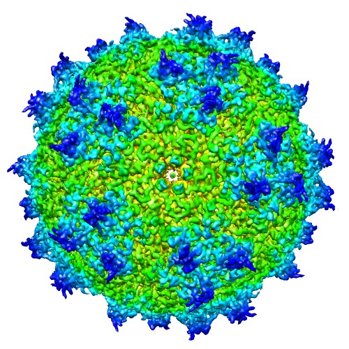 emd_5415_1.jpg emd_5415_1.jpg | 116 KB | ||
| Archive directory |  http://ftp.pdbj.org/pub/emdb/structures/EMD-5415ftp://ftp.pdbj.org/pub/emdb/structures/EMD-5415 http://ftp.pdbj.org/pub/emdb/structures/EMD-5415ftp://ftp.pdbj.org/pub/emdb/structures/EMD-5415 | HTTPS FTP |
-Related structure data
| Related structure data | 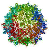 3j1qMC M: atomic model generated by this map C: citing same article ( |
|---|---|
| Similar structure data |


-Links
| EMDB pages | EMDB (EBI/PDBe) / EMDataResource |
|---|---|
| Related items in Molecule of the Month |


-Map
| File | Download / File: emd_5415.map.gz / Format: CCP4 / Size: 173.8 MB / Type: IMAGE STORED AS FLOATING POINT NUMBER (4 BYTES) | ||||||||||||||||||||||||||||||||||||||||||||||||||||||||||||||||||||
|---|---|---|---|---|---|---|---|---|---|---|---|---|---|---|---|---|---|---|---|---|---|---|---|---|---|---|---|---|---|---|---|---|---|---|---|---|---|---|---|---|---|---|---|---|---|---|---|---|---|---|---|---|---|---|---|---|---|---|---|---|---|---|---|---|---|---|---|---|---|
| Annotation | Reconstruction of AAV-DJ, a re-targeted gene therapy vector | ||||||||||||||||||||||||||||||||||||||||||||||||||||||||||||||||||||
| Voxel size | X=Y=Z: 1.3217 Å | ||||||||||||||||||||||||||||||||||||||||||||||||||||||||||||||||||||
| Density |
| ||||||||||||||||||||||||||||||||||||||||||||||||||||||||||||||||||||
| Symmetry | Space group: 1 | ||||||||||||||||||||||||||||||||||||||||||||||||||||||||||||||||||||
| Details | EMDB XML:
CCP4 map header:
| ||||||||||||||||||||||||||||||||||||||||||||||||||||||||||||||||||||
-Supplemental data
- Sample components
-Entire : AAV-DJ
| Entire | Name: AAV-DJ |
|---|---|
| Components |
|
-Supramolecule #1000: AAV-DJ
| Supramolecule | Name: AAV-DJ / type: sample / ID: 1000 Oligomeric state: 60 viral subunits form the icosahedral capsid Number unique components: 1 |
|---|---|
| Molecular weight | Theoretical: 3.75 MDa |
-Supramolecule #1: Adeno-associated virus
| Supramolecule | Name: Adeno-associated virus / type: virus / ID: 1 / NCBI-ID: 272636 / Sci species name: Adeno-associated virus / Database: NCBI / Virus type: VIRUS-LIKE PARTICLE / Virus isolate: SEROTYPE / Virus enveloped: No / Virus empty: Yes |
|---|---|
| Host (natural) | Organism:  Homo sapiens (human) / synonym: VERTEBRATES Homo sapiens (human) / synonym: VERTEBRATES |
| Molecular weight | Theoretical: 3.75 MDa |
| Virus shell | Shell ID: 1 / Name: VP3 / Diameter: 250 Å / T number (triangulation number): 1 |
-Experimental details
-Structure determination
| Method | cryo EM |
|---|---|
 Processing Processing | single particle reconstruction |
| Aggregation state | particle |
-Sample preparation
| Concentration | 1.5 mg/mL |
|---|---|
| Buffer | pH: 7.5 / Details: 125mM NaCl, 10mM Tris, 1mM MgCl2, pH 7.5 |
| Grid | Details: C-flat grid CF-2/1-2C (2um hole, 1 um spacing, 200 mesh copper), glow discharged |
| Vitrification | Cryogen name: ETHANE / Instrument: FEI VITROBOT MARK I |
- Electron microscopy
| Microscope | FEI TITAN KRIOS |
|---|---|
| Electron beam | Acceleration voltage: 300 kV / Electron source: FIELD EMISSION GUN |
| Electron optics | Illumination mode: FLOOD BEAM / Imaging mode: BRIGHT FIELDBright-field microscopy |
| Sample stage | Specimen holder model: FEI TITAN KRIOS AUTOGRID HOLDER |
| Date | Feb 25, 2011 |
| Image recording | Number real images: 4773 |
| Experimental equipment |  Model: Titan Krios / Image courtesy: FEI Company |
-Image processing
| CTF correction | Details: CTF was estimated using Appion software |
|---|---|
| Final reconstruction | Resolution.type: BY AUTHOR / Resolution: 4.5 Å / Resolution method: FSC 0.143 CUT-OFF / Software - Name: FREALIGN / Number images used: 27312 |
| Details | Images were collected automatically using Leginon software. Particles were manually selected for single particle reconstruction. |
-Atomic model buiding 1
| Initial model | PDB ID: Chain - Chain ID: A |
|---|---|
| Software | Name: RSRef, CNS |
| Details | Protocol: Refinement of reconstruction magnification relative to homolog crystal structure, manual rebuilding, simulated annealing torsion angle dynamics, isotropic B-factors. Iterative refinement in RSRef and model building in Coot |
| Refinement | Space: REAL / Protocol: FLEXIBLE FIT / Overall B value: 30 / Target criteria: Correlation coefficient |
| Output model | PDB-3j1q: |