 Movie
Movie Controller

[English] 日本語
 Yorodumi
- EMDB-5374: Structural basis for broad detection of genogroup II noroviruses ... -
+ Open data

- Basic information
| Entry | Database: EMDB / ID: EMD-5374 | |||||||||
|---|---|---|---|---|---|---|---|---|---|---|
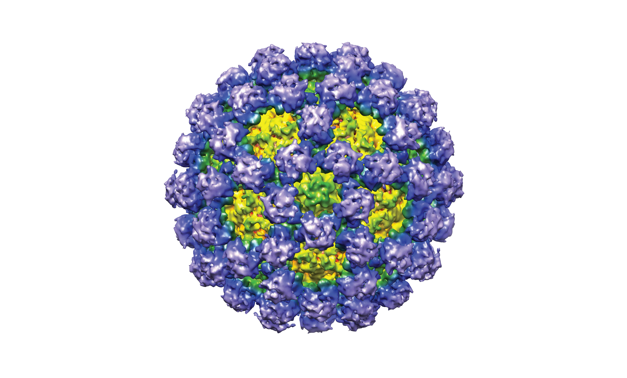
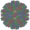
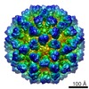
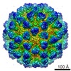
| Title | Structural basis for broad detection of genogroup II noroviruses by a monoclonal antibody that binds to a site occluded in the viral particle | |||||||||
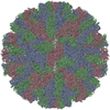
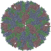
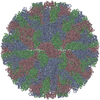
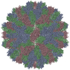
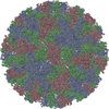
 Map data Map data | Surface rendering of GII.10 norovirus VLP | |||||||||
 Sample Sample |
| |||||||||
 Keywords Keywords |  norovirus norovirus | |||||||||
| Biological species |  Norovirus genogroup GII.10 Norovirus genogroup GII.10 | |||||||||
| Method | single particle reconstruction / cryo EM / Resolution: 10.0 Å | |||||||||
 Authors Authors | Hansman GS / Taylor DW / McLellan JS / Smith TJ / Georgiev I / Tame JRH / Park SY / Yamazaki M / Gondaira F / Miki M ...Hansman GS / Taylor DW / McLellan JS / Smith TJ / Georgiev I / Tame JRH / Park SY / Yamazaki M / Gondaira F / Miki M / Katayama K / Murata K / Kwong PD | |||||||||
 Citation Citation | Journal: J Virol / Year: 2012 Title: Structural basis for broad detection of genogroup II noroviruses by a monoclonal antibody that binds to a site occluded in the viral particle. Authors: Grant S Hansman / David W Taylor / Jason S McLellan / Thomas J Smith / Ivelin Georgiev / Jeremy R H Tame / Sam-Yong Park / Makoto Yamazaki / Fumio Gondaira / Motohiro Miki / Kazuhiko ...Authors: Grant S Hansman / David W Taylor / Jason S McLellan / Thomas J Smith / Ivelin Georgiev / Jeremy R H Tame / Sam-Yong Park / Makoto Yamazaki / Fumio Gondaira / Motohiro Miki / Kazuhiko Katayama / Kazuyoshi Murata / Peter D Kwong /  Abstract: Human noroviruses are genetically and antigenically highly divergent. Monoclonal antibodies raised in mice against one kind of norovirus virus-like particle (VLP), however, were found to have broad ...Human noroviruses are genetically and antigenically highly divergent. Monoclonal antibodies raised in mice against one kind of norovirus virus-like particle (VLP), however, were found to have broad recognition. In this study, we present the crystal structure of the antigen-binding fragment (Fab) for one of these broadly reactive monoclonal antibodies, 5B18, in complex with the capsid-protruding domain from a genogroup II genotype 10 (GII.10) norovirus at 3.3-Å resolution and, also, the cryo-electron microscopy structure of the GII.10 VLP at ∼10-Å resolution. The GII.10 VLP structure was more similar in overall architecture to the GV.1 murine norovirus virion than to the prototype GI.1 human norovirus VLP, with the GII.10 protruding domain raised ∼15 Å off the shell domain and rotated ∼40° relative to the GI.1 protruding domain. In the crystal structure, the 5B18 Fab bound to a highly conserved region of the protruding domain. Based on the VLP structure, this region is involved in interactions with other regions of the capsid and is buried in the virus particle. Despite the occluded nature of the recognized epitope in the VLP structure, enzyme-linked immunosorbent assay (ELISA) binding suggested that the 5B18 antibody was able to capture intact VLPs. Together, the results provide evidence that the norovirus particle is capable of extreme conformational flexibility, which may allow for antibody recognition of conserved surfaces that would otherwise be buried on intact particles. | |||||||||
| History |
|
- Structure visualization
| Movie |
 Movie viewer Movie viewer |
|---|---|
| Structure viewer | EM map: SurfViewMolmilJmol/JSmol |
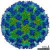
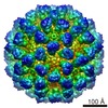
| Supplemental images |
- Downloads & links
-EMDB archive
| Map data | emd_5374.map.gz | 54.1 MB | EMDB map data format | |
|---|---|---|---|---|
| Header (meta data) | emd-5374-v30.xmlemd-5374.xml | 10 KB 10 KB | Display Display | EMDB header |
| Images |  emd_5374.png emd_5374.png | 1.5 MB | ||
| Archive directory |  http://ftp.pdbj.org/pub/emdb/structures/EMD-5374ftp://ftp.pdbj.org/pub/emdb/structures/EMD-5374 http://ftp.pdbj.org/pub/emdb/structures/EMD-5374ftp://ftp.pdbj.org/pub/emdb/structures/EMD-5374 | HTTPS FTP |
-Related structure data

-Links
| EMDB pages | EMDB (EBI/PDBe) / EMDataResource |
|---|
-Map
| File | Download / File: emd_5374.map.gz / Format: CCP4 / Size: 58.2 MB / Type: IMAGE STORED AS FLOATING POINT NUMBER (4 BYTES) | ||||||||||||||||||||||||||||||||||||||||||||||||||||||||||||||||||||
|---|---|---|---|---|---|---|---|---|---|---|---|---|---|---|---|---|---|---|---|---|---|---|---|---|---|---|---|---|---|---|---|---|---|---|---|---|---|---|---|---|---|---|---|---|---|---|---|---|---|---|---|---|---|---|---|---|---|---|---|---|---|---|---|---|---|---|---|---|---|
| Annotation | Surface rendering of GII.10 norovirus VLP | ||||||||||||||||||||||||||||||||||||||||||||||||||||||||||||||||||||
| Voxel size | X=Y=Z: 2.4 Å | ||||||||||||||||||||||||||||||||||||||||||||||||||||||||||||||||||||
| Density |
| ||||||||||||||||||||||||||||||||||||||||||||||||||||||||||||||||||||
| Symmetry | Space group: 1 | ||||||||||||||||||||||||||||||||||||||||||||||||||||||||||||||||||||
| Details | EMDB XML:
CCP4 map header:
| ||||||||||||||||||||||||||||||||||||||||||||||||||||||||||||||||||||
-Supplemental data
- Sample components
-Entire : GII.10 norovirus VLP
| Entire | Name: GII.10 norovirus VLP |
|---|---|
| Components |
|
-Supramolecule #1000: GII.10 norovirus VLP
| Supramolecule | Name: GII.10 norovirus VLP / type: sample / ID: 1000 / Oligomeric state: icosahedral (T3) / Number unique components: 1 |
|---|---|
| Molecular weight | Experimental: 10 MDa / Theoretical: 10 MDa |
-Supramolecule #1: Norovirus genogroup GII.10
| Supramolecule | Name: Norovirus genogroup GII.10 / type: virus / ID: 1 / Name.synonym: GII.10 Vietnam 026 VLP / NCBI-ID: 747305 / Sci species name: Norovirus genogroup GII.10 / Database: NCBI / Virus type: VIRUS-LIKE PARTICLE / Virus isolate: STRAIN / Virus enveloped: No / Virus empty: Yes / Syn species name: GII.10 Vietnam 026 VLP |
|---|---|
| Host (natural) | Organism:  Homo sapiens (human) / synonym: VERTEBRATES Homo sapiens (human) / synonym: VERTEBRATES |
| Molecular weight | Experimental: 10 MDa / Theoretical: 10 MDa |
| Virus shell | Shell ID: 1 / T number (triangulation number): 3 |
-Experimental details
-Structure determination
| Method | cryo EM |
|---|---|
 Processing Processing | single particle reconstruction |
| Aggregation state | particle |
-Sample preparation
| Concentration | 1.0 mg/mL |
|---|---|
| Buffer | pH: 7.3 / Details: PBS |
| Grid | Details: Quantifoil R1.2/1.3 Mo 200 mesh |
| Vitrification | Cryogen name: ETHANE / Chamber humidity: 100 % / Instrument: FEI VITROBOT MARK IV Details: Vitrification instrument: FEI Vitrobot Mark IV. Quantifoil Mo 200 mesh holey carbon grid with a thin layer of carbon over the holes Method: automatic blotting for 8 seconds |
- Electron microscopy
| Microscope | JEOL 2200FS |
|---|---|
| Electron beam | Acceleration voltage: 200 kV / Electron source: FIELD EMISSION GUN |
| Electron optics | Illumination mode: FLOOD BEAM / Imaging mode: BRIGHT FIELDBright-field microscopy / Nominal defocus max: 2.0 µm / Nominal defocus min: 0.5 µm / Nominal magnification: 40000 |
| Sample stage | Specimen holder: Gatan / Specimen holder model: GATAN LIQUID NITROGEN |
| Date | Jul 22, 2011 |
| Image recording | Category: CCD / Film or detector model: GENERIC TVIPS (4k x 4k) / Average electron dose: 20 e/Å2 |
-Image processing
| CTF correction | Details: Each image |
|---|---|
| Final reconstruction | Algorithm: OTHER / Resolution.type: BY AUTHOR / Resolution: 10.0 Å / Resolution method: FSC 0.5 CUT-OFF / Software - Name: EMAN2 / Number images used: 8000 |
| Details | The particles were selected semi-automatically using the swarm program in EMAN2. |
