 Movie
Movie Controller

[English] 日本語
 Yorodumi
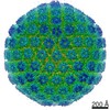
- EMDB-5255: Seeing the Portal in Herpes Simplex Virus type I B-capsids. Map1:... -
+ Open data

- Basic information
| Entry | Database: EMDB / ID: EMD-5255 | |||||||||
|---|---|---|---|---|---|---|---|---|---|---|
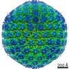
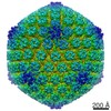
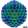
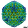
| Title | Seeing the Portal in Herpes Simplex Virus type I B-capsids. Map1: Icosahedral reconstruction of the HSV-1 B-capsid Map2: symmetry free (c1) reconstruction of the HSV-1 B-capsid Map3: c12 symmetrized portal density extracted from symmetry free (c1) reconstruction of the HSV-1 B-capsid Map4: c1 portal density extracted from symmetry free (c1) reconstruction of the HSV-1 B-capsid | |||||||||
 Map data Map data | This is the non-averaged portal density extracted from the c1 (symmetry-free) reconstruction of the HSV-1 B-capsid | |||||||||
 Sample Sample |
| |||||||||
 Keywords Keywords | Herpes / HSV1 / Portal / Cryo-EM / Single Particle / UL6 / Asymmetric / Symmetry-free | |||||||||
| Biological species |   Herpes Simplex Virus Type I (Herpes simplex virus type 1) Herpes Simplex Virus Type I (Herpes simplex virus type 1) | |||||||||
| Method | single particle reconstruction / cryo EM / Resolution: 24.0 Å | |||||||||
 Authors Authors | Rochat RH / Liu X / Murata K / Nagayama K / Rixon FJ / Chiu W | |||||||||
 Citation Citation | Journal: J Virol / Year: 2011 Title: Seeing the portal in herpes simplex virus type 1 B capsids. Authors: R H Rochat / X Liu / K Murata / K Nagayama / F J Rixon / W Chiu /  Abstract: Resolving the nonicosahedral components in large icosahedral viruses remains a technical challenge in structural virology. We have used the emerging technique of Zernike phase-contrast electron ...Resolving the nonicosahedral components in large icosahedral viruses remains a technical challenge in structural virology. We have used the emerging technique of Zernike phase-contrast electron cryomicroscopy to enhance the image contrast of ice-embedded herpes simplex virus type 1 capsids. Image reconstruction enabled us to retrieve the structure of the unique portal vertex in the context of the icosahedral capsid and, for the first time, show the subunit organization of a portal in a virus infecting eukaryotes. Our map unequivocally resolves the 12-subunit portal situated beneath one of the pentameric vertices, thus removing uncertainty over the location and stoichiometry of the herpesvirus portal. | |||||||||
| History |
|
- Structure visualization
| Movie |
 Movie viewer Movie viewer |
|---|---|
| Structure viewer | EM map: SurfViewMolmilJmol/JSmol |
| Supplemental images |

- Downloads & links
-EMDB archive
| Map data | emd_5255.map.gz | 27.7 MB | EMDB map data format | |
|---|---|---|---|---|
| Header (meta data) | emd-5255-v30.xmlemd-5255.xml | 11.1 KB 11.1 KB | Display Display | EMDB header |
| Images | 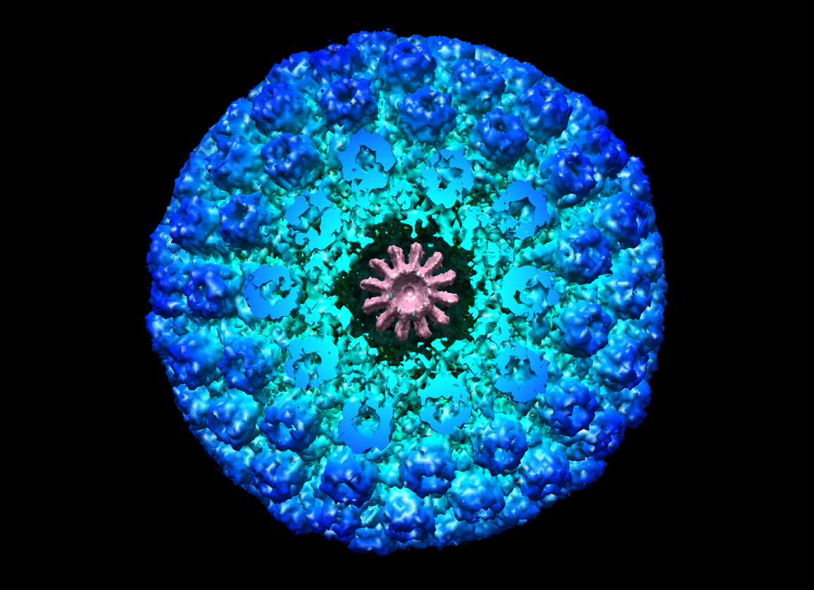 emd_5255_1.png emd_5255_1.png | 725.2 KB | ||
| Archive directory |  http://ftp.pdbj.org/pub/emdb/structures/EMD-5255ftp://ftp.pdbj.org/pub/emdb/structures/EMD-5255 http://ftp.pdbj.org/pub/emdb/structures/EMD-5255ftp://ftp.pdbj.org/pub/emdb/structures/EMD-5255 | HTTPS FTP |
-Validation report
| Summary document | emd_5255_validation.pdf.gz | 78.9 KB | Display | EMDB validaton report |
|---|---|---|---|---|
| Full document | emd_5255_full_validation.pdf.gz | 78 KB | Display | |
| Data in XML | emd_5255_validation.xml.gz | 493 B | Display | |
| Arichive directory | https://ftp.pdbj.org/pub/emdb/validation_reports/EMD-5255ftp://ftp.pdbj.org/pub/emdb/validation_reports/EMD-5255 | HTTPS FTP |
-Related structure data
-Links
| EMDB pages | EMDB (EBI/PDBe) / EMDataResource |
|---|
-Map
| File | Download / File: emd_5255.map.gz / Format: CCP4 / Size: 100.6 MB / Type: IMAGE STORED AS FLOATING POINT NUMBER (4 BYTES) | ||||||||||||||||||||||||||||||||||||||||||||||||||||||||||||||||||||
|---|---|---|---|---|---|---|---|---|---|---|---|---|---|---|---|---|---|---|---|---|---|---|---|---|---|---|---|---|---|---|---|---|---|---|---|---|---|---|---|---|---|---|---|---|---|---|---|---|---|---|---|---|---|---|---|---|---|---|---|---|---|---|---|---|---|---|---|---|---|
| Annotation | This is the non-averaged portal density extracted from the c1 (symmetry-free) reconstruction of the HSV-1 B-capsid | ||||||||||||||||||||||||||||||||||||||||||||||||||||||||||||||||||||
| Voxel size | X=Y=Z: 5.32 Å | ||||||||||||||||||||||||||||||||||||||||||||||||||||||||||||||||||||
| Density |
| ||||||||||||||||||||||||||||||||||||||||||||||||||||||||||||||||||||
| Symmetry | Space group: 1 | ||||||||||||||||||||||||||||||||||||||||||||||||||||||||||||||||||||
| Details | EMDB XML:
CCP4 map header:
| ||||||||||||||||||||||||||||||||||||||||||||||||||||||||||||||||||||
-Supplemental data
- Sample components
-Entire : Herpes simplex virus type I B-capsids
| Entire | Name: Herpes simplex virus type I B-capsids |
|---|---|
| Components |
|
-Supramolecule #1000: Herpes simplex virus type I B-capsids
| Supramolecule | Name: Herpes simplex virus type I B-capsids / type: sample / ID: 1000 / Oligomeric state: Icosahedral / Number unique components: 1 |
|---|---|
| Molecular weight | Theoretical: 100 MDa |
-Supramolecule #1: Herpes Simplex Virus Type I
| Supramolecule | Name: Herpes Simplex Virus Type I / type: virus / ID: 1 / Name.synonym: HSV-1 / Sci species name: Herpes Simplex Virus Type I / Database: NCBI / Virus type: OTHER / Virus isolate: STRAIN / Virus enveloped: No / Virus empty: No / Syn species name: HSV-1 |
|---|---|
| Host (natural) | Organism:  Homo sapiens (human) / synonym: VERTEBRATES Homo sapiens (human) / synonym: VERTEBRATES |
| Molecular weight | Theoretical: 200 MDa |
| Virus shell | Shell ID: 1 / Name: vp5 / Diameter: 1250 Å / T number (triangulation number): 16 |
| Virus shell | Shell ID: 2 / Name: vp26 / Diameter: 1250 Å / T number (triangulation number): 15 |
| Virus shell | Shell ID: 3 / Name: vp19 / Diameter: 1250 Å / T number (triangulation number): 10 |
| Virus shell | Shell ID: 4 / Name: vp23 / Diameter: 1250 Å / T number (triangulation number): 5 |
-Experimental details
-Structure determination
| Method | cryo EM |
|---|---|
 Processing Processing | single particle reconstruction |
| Aggregation state | particle |
-Sample preparation
| Grid | Details: Quantifoil 2/2 grids |
|---|---|
| Vitrification | Cryogen name: ETHANE / Chamber humidity: 100 % / Chamber temperature: 85 K / Instrument: FEI VITROBOT MARK IV / Details: Vitrification instrument: Vitrobot Mark IV / Method: Blot 1 time for 2 seconds |
- Electron microscopy
| Microscope | JEOL 2200FS |
|---|---|
| Temperature | Min: 100 K / Max: 101 K / Average: 100 K |
| Alignment procedure | Legacy - Astigmatism: Astigmatism was corrected at 400,000 times magnification |
| Specialist optics | Energy filter - Name: Omega / Energy filter - Lower energy threshold: 0.0 eV / Energy filter - Upper energy threshold: 20.0 eV |
| Date | Jun 10, 2009 |
| Image recording | Category: CCD / Film or detector model: GENERIC TVIPS (4k x 4k) / Average electron dose: 20 e/Å2 |
| Tilt angle min | 0 |
| Tilt angle max | 0 |
| Electron beam | Acceleration voltage: 200 kV / Electron source:  FIELD EMISSION GUN FIELD EMISSION GUN |
| Electron optics | Calibrated magnification: 56000 / Illumination mode: FLOOD BEAM / Imaging mode: BRIGHT FIELD / Cs: 4.1 mm / Nominal defocus max: 0.2 µm / Nominal defocus min: 0.0 µm / Nominal magnification: 40000 |
| Sample stage | Specimen holder: Side Entry / Specimen holder model: GATAN LIQUID NITROGEN |
-Image processing
| Details | The particles were aligned using the multi-path simulated annealing algorithm. |
|---|---|
| CTF correction | Details: No Correction |
| Final reconstruction | Algorithm: OTHER / Resolution.type: BY AUTHOR / Resolution: 24.0 Å / Resolution method: FSC 0.5 CUT-OFF / Software - Name: MPSA Details: Reconstructions were made both with and without imposed symmetry. Number images used: 6300 |
