 Movie
Movie Controller


+ Open data

- Basic information
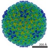
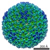
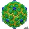
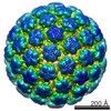
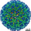
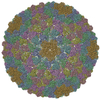
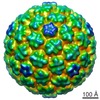
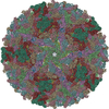
| Entry | Database: EMDB / ID: EMD-5149 | |||||||||
|---|---|---|---|---|---|---|---|---|---|---|
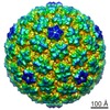
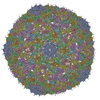
| Title | P22 Procapsid Shell | |||||||||
 Map data Map data | This is a density map of WT P22 procapsid shells (devoid of scaffolding protein) | |||||||||
 Sample Sample |
| |||||||||
 Keywords Keywords | Precursor capsid structure for bacteriophage P22 | |||||||||
| Function / homology | Major capsid protein Gp5 / P22 coat protein - gene protein 5 / viral procapsid / viral procapsid maturation / T=7 icosahedral viral capsid /  viral capsid / identical protein binding / Major capsid protein viral capsid / identical protein binding / Major capsid protein Function and homology information Function and homology information | |||||||||
| Biological species |  Enterobacteria phage P22 (virus) Enterobacteria phage P22 (virus) | |||||||||
| Method | single particle reconstruction / cryo EM / Resolution: 9.1 Å | |||||||||
 Authors Authors | Parent KN / Khayat R / Tu LH / Suhanovsky MM / Coritnes JR / Teschke CM / Johnson JE / Baker TS | |||||||||
 Citation Citation | Journal: Structure / Year: 2010 Title: P22 coat protein structures reveal a novel mechanism for capsid maturation: stability without auxiliary proteins or chemical crosslinks. Authors: Kristin N Parent / Reza Khayat / Long H Tu / Margaret M Suhanovsky / Juliana R Cortines / Carolyn M Teschke / John E Johnson / Timothy S Baker /  Abstract: Viral capsid assembly and stability in tailed, dsDNA phage and Herpesviridae are achieved by various means including chemical crosslinks (unique to HK97), or auxiliary proteins (lambda, T4, phi29, ...Viral capsid assembly and stability in tailed, dsDNA phage and Herpesviridae are achieved by various means including chemical crosslinks (unique to HK97), or auxiliary proteins (lambda, T4, phi29, and herpesviruses). All these viruses have coat proteins (CP) with a conserved, HK97-like core structure. We used a combination of trypsin digestion, gold labeling, cryo-electron microscopy, 3D image reconstruction, and comparative modeling to derive two independent, pseudoatomic models of bacteriophage P22 CP: before and after maturation. P22 capsid stabilization results from intersubunit interactions among N-terminal helices and an extensive "P loop," which obviate the need for crosslinks or auxiliary proteins. P22 CP also has a telokin-like Ig domain that likely stabilizes the monomer fold so that assembly may proceed via individual subunit addition rather than via preformed capsomers as occurs in HK97. Hence, the P22 CP structure may be a paradigm for understanding how monomers assemble in viruses like phi29 and HSV-1. | |||||||||
| History |
|
- Structure visualization
| Movie |
Movie viewer |
|---|---|
| Structure viewer | EM map: SurfViewMolmilJmol/JSmol |
| Supplemental images |

- Downloads & links
-EMDB archive
| Map data | emd_5149.map.gz | 152.1 MB | EMDB map data format | |
|---|---|---|---|---|
| Header (meta data) | emd-5149-v30.xmlemd-5149.xml | 10.4 KB 10.4 KB | Display Display | EMDB header |
| Images | emd_5149_1.tif | 761.4 KB | ||
| Archive directory |  http://ftp.pdbj.org/pub/emdb/structures/EMD-5149ftp://ftp.pdbj.org/pub/emdb/structures/EMD-5149 http://ftp.pdbj.org/pub/emdb/structures/EMD-5149ftp://ftp.pdbj.org/pub/emdb/structures/EMD-5149 | HTTPS FTP |
-Related structure data
| Related structure data | 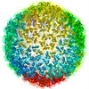 3iyiMC  5150C 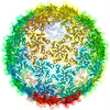 3iyhC M: atomic model generated by this map C: citing same article ( |
|---|---|
| Similar structure data |
-Links
| EMDB pages | EMDB (EBI/PDBe) / EMDataResource |
|---|---|
| Related items in Molecule of the Month |

-Map
| File | Download / File: emd_5149.map.gz / Format: CCP4 / Size: 296.2 MB / Type: IMAGE STORED AS FLOATING POINT NUMBER (4 BYTES) | ||||||||||||||||||||||||||||||||||||||||||||||||||||||||||||||||||||
|---|---|---|---|---|---|---|---|---|---|---|---|---|---|---|---|---|---|---|---|---|---|---|---|---|---|---|---|---|---|---|---|---|---|---|---|---|---|---|---|---|---|---|---|---|---|---|---|---|---|---|---|---|---|---|---|---|---|---|---|---|---|---|---|---|---|---|---|---|---|
| Annotation | This is a density map of WT P22 procapsid shells (devoid of scaffolding protein) | ||||||||||||||||||||||||||||||||||||||||||||||||||||||||||||||||||||
| Voxel size | X=Y=Z: 1.62 Å | ||||||||||||||||||||||||||||||||||||||||||||||||||||||||||||||||||||
| Density |
| ||||||||||||||||||||||||||||||||||||||||||||||||||||||||||||||||||||
| Symmetry | Space group: 1 | ||||||||||||||||||||||||||||||||||||||||||||||||||||||||||||||||||||
| Details | EMDB XML:
CCP4 map header:
| ||||||||||||||||||||||||||||||||||||||||||||||||||||||||||||||||||||
-Supplemental data
- Sample components
-Entire : P22 Procapsid Shell
| Entire | Name: P22 Procapsid Shell |
|---|---|
| Components |
|
-Supramolecule #1000: P22 Procapsid Shell
| Supramolecule | Name: P22 Procapsid Shell / type: sample / ID: 1000 / Oligomeric state: Icosahedral / Number unique components: 1 |
|---|
-Supramolecule #1: Enterobacteria phage P22
| Supramolecule | Name: Enterobacteria phage P22 / type: virus / ID: 1 / Name.synonym: Phage P22 / NCBI-ID: 10754 / Sci species name: Enterobacteria phage P22 / Database: NCBI / Virus type: OTHER / Virus isolate: STRAIN / Virus enveloped: No / Virus empty: Yes / Syn species name: Phage P22 |
|---|---|
| Host (natural) | Organism:  Salmonella enterica subsp. enterica serovar Typhimurium (bacteria) Salmonella enterica subsp. enterica serovar Typhimurium (bacteria)synonym: BACTERIA(EUBACTERIA) |
| Virus shell | Shell ID: 1 / Name: P22 Procapsid shell / Diameter: 580 Å / T number (triangulation number): 7 |
-Experimental details
-Structure determination
| Method | cryo EM |
|---|---|
 Processing Processing | single particle reconstruction |
| Aggregation state | particle |
-Sample preparation
| Concentration | 8 mg/mL |
|---|---|
| Buffer | pH: 7.6 / Details: 20 mM sodium phosphate |
| Grid | Details: Quantifoil grids |
| Vitrification | Cryogen name: ETHANE / Chamber humidity: 100 % / Chamber temperature: 89 K / Instrument: HOMEMADE PLUNGER / Details: Vitrification instrument: manual plunge-freezer / Method: Blot for 2-3 seconds before plunging |
- Electron microscopy
| Microscope | FEI POLARA 300 |
|---|---|
| Electron beam | Acceleration voltage: 200 kV / Electron source: FIELD EMISSION GUN |
| Electron optics | Illumination mode: FLOOD BEAM / Imaging mode: BRIGHT FIELDBright-field microscopy / Cs: 2.3 mm / Nominal defocus max: 3.65 µm / Nominal defocus min: 0.41 µm / Nominal magnification: 39000 |
| Sample stage | Specimen holder: Polrar Multi Specimen Holder / Specimen holder model: OTHER |
| Temperature | Min: 90 K / Max: 90 K / Average: 90 K |
| Alignment procedure | Legacy - Astigmatism: at working magnification |
| Date | Aug 10, 2009 |
| Image recording | Category: FILM / Film or detector model: KODAK SO-163 FILM / Digitization - Scanner: OTHER / Digitization - Sampling interval: 6.35 µm / Number real images: 164 / Average electron dose: 8 e/Å2 / Details: scanned on Nikon SuperCool 8000 / Bits/pixel: 8 |
| Tilt angle min | 0 |
| Tilt angle max | 0 |
| Experimental equipment |  Model: Tecnai Polara / Image courtesy: FEI Company |
-Image processing
| CTF correction | Details: ROBEM |
|---|---|
| Final reconstruction | Algorithm: OTHER / Resolution.type: BY AUTHOR / Resolution: 9.1 Å / Resolution method: FSC 0.5 CUT-OFF / Software - Name: AUTO3DEM / Number images used: 11997 |
| Details | data were collected on film |
