 Movie
Movie Controller


+ Open data

- Basic information
| Entry | Database: EMDB / ID: EMD-3451 | |||||||||
|---|---|---|---|---|---|---|---|---|---|---|
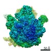
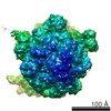
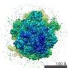
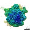
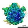
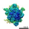
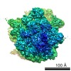
| Title | Folding intermediate of spectrin R16 bound to 70s ribosome | |||||||||
 Map data Map data | Folding intermediate of spectrin R16 bound to 70s ribosome | |||||||||
 Sample Sample |
| |||||||||
| Function / homology |  Function and homology information Function and homology information costamere / actin filament capping / cortical actin cytoskeleton / cell projection / actin filament binding / cell junction / actin cytoskeleton organization / calmodulin binding / calcium ion binding / plasma membrane costamere / actin filament capping / cortical actin cytoskeleton / cell projection / actin filament binding / cell junction / actin cytoskeleton organization / calmodulin binding / calcium ion binding / plasma membraneSimilarity search - Function | |||||||||
| Biological species |  Escherichia coli (E. coli) Escherichia coli (E. coli) | |||||||||
| Method | single particle reconstruction / cryo EM / Resolution: 4.8 Å | |||||||||
 Authors Authors | Nilsson OB / Nickson AA / Clarke J | |||||||||
 Citation Citation | Journal: Nat Struct Mol Biol / Year: 2017 Title: Cotranslational folding of spectrin domains via partially structured states. Authors: Ola B Nilsson / Adrian A Nickson / Jeffrey J Hollins / Stephan Wickles / Annette Steward / Roland Beckmann / Gunnar von Heijne / Jane Clarke /    Abstract: How do the key features of protein folding, elucidated from studies on native, isolated proteins, manifest in cotranslational folding on the ribosome? Using a well-characterized family of homologous ...How do the key features of protein folding, elucidated from studies on native, isolated proteins, manifest in cotranslational folding on the ribosome? Using a well-characterized family of homologous α-helical proteins with a range of biophysical properties, we show that spectrin domains can fold vectorially on the ribosome and may do so via a pathway different from that of the isolated domain. We use cryo-EM to reveal a folded or partially folded structure, formed in the vestibule of the ribosome. Our results reveal that it is not possible to predict which domains will fold within the ribosome on the basis of the folding behavior of isolated domains; instead, we propose that a complex balance of the rate of folding, the rate of translation and the lifetime of folded or partly folded states will determine whether folding occurs cotranslationally on actively translating ribosomes. | |||||||||
| History |
|
- Structure visualization
| Movie |
Movie viewer |
|---|---|
| Structure viewer | EM map: SurfViewMolmilJmol/JSmol |
| Supplemental images |

- Downloads & links
-EMDB archive
| Map data | emd_3451.map.gz | 177.3 MB | EMDB map data format | |
|---|---|---|---|---|
| Header (meta data) | emd-3451-v30.xmlemd-3451.xml | 12.2 KB 12.2 KB | Display Display | EMDB header |
| Images | 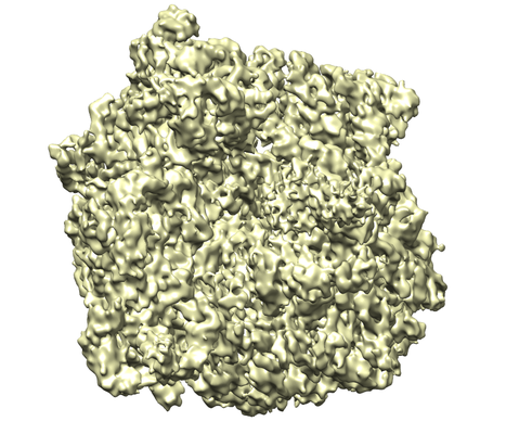 emd_3451.png emd_3451.png | 224.7 KB | ||
| Archive directory |  http://ftp.pdbj.org/pub/emdb/structures/EMD-3451ftp://ftp.pdbj.org/pub/emdb/structures/EMD-3451 http://ftp.pdbj.org/pub/emdb/structures/EMD-3451ftp://ftp.pdbj.org/pub/emdb/structures/EMD-3451 | HTTPS FTP |
-Related structure data
| Related structure data |  5m6sMC M: atomic model generated by this map C: citing same article ( |
|---|---|
| Similar structure data |
-Links
| EMDB pages | EMDB (EBI/PDBe) / EMDataResource |
|---|---|
| Related items in Molecule of the Month |

-Map
| File | Download / File: emd_3451.map.gz / Format: CCP4 / Size: 190.1 MB / Type: IMAGE STORED AS FLOATING POINT NUMBER (4 BYTES) | ||||||||||||||||||||||||||||||||||||||||||||||||||||||||||||
|---|---|---|---|---|---|---|---|---|---|---|---|---|---|---|---|---|---|---|---|---|---|---|---|---|---|---|---|---|---|---|---|---|---|---|---|---|---|---|---|---|---|---|---|---|---|---|---|---|---|---|---|---|---|---|---|---|---|---|---|---|---|
| Annotation | Folding intermediate of spectrin R16 bound to 70s ribosome | ||||||||||||||||||||||||||||||||||||||||||||||||||||||||||||
| Voxel size | X=Y=Z: 1.084 Å | ||||||||||||||||||||||||||||||||||||||||||||||||||||||||||||
| Density |
| ||||||||||||||||||||||||||||||||||||||||||||||||||||||||||||
| Symmetry | Space group: 1 | ||||||||||||||||||||||||||||||||||||||||||||||||||||||||||||
| Details | EMDB XML:
CCP4 map header:
| ||||||||||||||||||||||||||||||||||||||||||||||||||||||||||||
-Supplemental data
- Sample components
-Entire : Folding intermediate of spectrin R16 bound to 70s ribosome
| Entire | Name: Folding intermediate of spectrin R16 bound to 70s ribosome |
|---|---|
| Components |
|
-Supramolecule #1: Folding intermediate of spectrin R16 bound to 70s ribosome
| Supramolecule | Name: Folding intermediate of spectrin R16 bound to 70s ribosome type: complex / ID: 1 / Parent: 0 |
|---|---|
| Source (natural) | Organism: Escherichia coli (E. coli) |
| Molecular weight | Theoretical: 2.5 MDa |
-Experimental details
-Structure determination
| Method | cryo EM |
|---|---|
 Processing Processing | single particle reconstruction |
| Aggregation state | particle |
-Sample preparation
| Buffer | pH: 7.4 Component:
| ||||||||||||
|---|---|---|---|---|---|---|---|---|---|---|---|---|---|
| Grid | Model: Quantifoil R3/3 / Material: COPPER/PALLADIUM / Support film - Material: CARBON / Support film - topology: CONTINUOUS / Support film - Film thickness: 2.0 nm / Pretreatment - Type: GLOW DISCHARGE | ||||||||||||
| Vitrification | Cryogen name: ETHANE / Chamber humidity: 100 % / Chamber temperature: 278 K / Instrument: FEI VITROBOT MARK IV | ||||||||||||
| Details | Folding intermediate of spectrin R16 bound to 70s ribosome |
- Electron microscopy
| Microscope | FEI TITAN |
|---|---|
| Electron beam | Acceleration voltage: 300 kV / Electron source: FIELD EMISSION GUN |
| Electron optics | Calibrated defocus min: 0.8 µm / Illumination mode: FLOOD BEAM / Imaging mode: BRIGHT FIELDBright-field microscopy / Cs: 2.7 mm / Nominal defocus max: 2.5 µm |
| Sample stage | Specimen holder model: FEI TITAN KRIOS AUTOGRID HOLDER / Cooling holder cryogen: NITROGEN |
| Image recording | #0 - Image recording ID: 1 / #0 - Film or detector model: FEI FALCON II (4k x 4k) / #0 - Average electron dose: 2.4 e/Å2 / #1 - Image recording ID: 2 / #1 - Film or detector model: FEI FALCON II (4k x 4k) / #1 - Average electron dose: 2.4 e/Å2 |
-Image processing
| CTF correction | Software - Name: CTFFIND (ver. 4) Details: CTF correction was done following 3D reconstruction. |
|---|---|
| Startup model | Type of model: OTHER / Details: 70S ribosome |
| Initial angle assignment | Type: PROJECTION MATCHING Projection matching processing - Number reference projections: 83 Software - Name: SPIDER (ver. 09.03) |
| Final 3D classification | Software - Name: SPIDER (ver. 09.03) |
| Final angle assignment | Type: PROJECTION MATCHING / Software - Name: SPIDER (ver. 09.03) |
| Final reconstruction | Resolution.type: BY AUTHOR / Resolution: 4.8 Å / Resolution method: FSC 0.143 CUT-OFF / Software - Name: SPIDER (ver. 09.03) Details: To exclude potential overfitting, the data were processed using a frequency limited refinement protocol by truncating high frequencies (low-pass filter at 8A) Number images used: 46067 |
| Image recording ID | 1 |
-Atomic model buiding 1
| Refinement | Protocol: BACKBONE TRACE |
|---|---|
| Output model | PDB-5m6s: |
