 Movie
Movie Controller


+ Open data

- Basic information
| Entry | Database: EMDB / ID: EMD-3204 | |||||||||
|---|---|---|---|---|---|---|---|---|---|---|
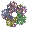
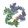
| Title | Structures of E.coli lysine decarboxylases | |||||||||
 Map data Map data | Reconstruction of active induced Lysine decarboxylase from E.coli | |||||||||
 Sample Sample |
| |||||||||
 Keywords Keywords | acid-stress /  lysine decarboxylase / RavA / cage lysine decarboxylase / RavA / cage | |||||||||
| Function / homology |  Function and homology information Function and homology informationlysine catabolic process / lysine decarboxylase / lysine decarboxylase activity / guanosine tetraphosphate binding / identical protein binding / cytoplasmSimilarity search - Function | |||||||||
| Biological species |  Escherichia coli K-12 (bacteria) Escherichia coli K-12 (bacteria) | |||||||||
| Method | single particle reconstruction / cryo EM / Resolution: 6.1 Å | |||||||||
 Authors Authors | Kandiah E / Carriel D / Perard J / Malet H / Bacia M / Liu K / Chan WSS / Houry AW / Ollagnier de Choudens S / Elsen S / Gutsche I | |||||||||
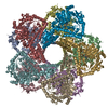
 Citation Citation | Journal: Elife / Year: 2014 Title: Assembly principles of a unique cage formed by hexameric and decameric E. coli proteins. Authors: Hélène Malet / Kaiyin Liu / Majida El Bakkouri / Sze Wah Samuel Chan / Gregory Effantin / Maria Bacia / Walid A Houry / Irina Gutsche /   Abstract: A 3.3 MDa macromolecular cage between two Escherichia coli proteins with seemingly incompatible symmetries-the hexameric AAA+ ATPase RavA and the decameric inducible lysine decarboxylase LdcI-is ...A 3.3 MDa macromolecular cage between two Escherichia coli proteins with seemingly incompatible symmetries-the hexameric AAA+ ATPase RavA and the decameric inducible lysine decarboxylase LdcI-is reconstructed by cryo-electron microscopy to 11 Å resolution. Combined with a 7.5 Å resolution reconstruction of the minimal complex between LdcI and the LdcI-binding domain of RavA, and the previously solved crystal structures of the individual components, this work enables to build a reliable pseudoatomic model of this unusual architecture and to identify conformational rearrangements and specific elements essential for complex formation. The design of the cage created via lateral interactions between five RavA rings is unique for the diverse AAA+ ATPase superfamily. | |||||||||
| History |
|
- Structure visualization
| Movie |
Movie viewer |
|---|---|
| Structure viewer | EM map: SurfViewMolmilJmol/JSmol |
| Supplemental images |

- Downloads & links
-EMDB archive
| Map data | emd_3204.map.gz | 60 MB | EMDB map data format | |
|---|---|---|---|---|
| Header (meta data) | emd-3204-v30.xmlemd-3204.xml | 11.5 KB 11.5 KB | Display Display | EMDB header |
| Images | 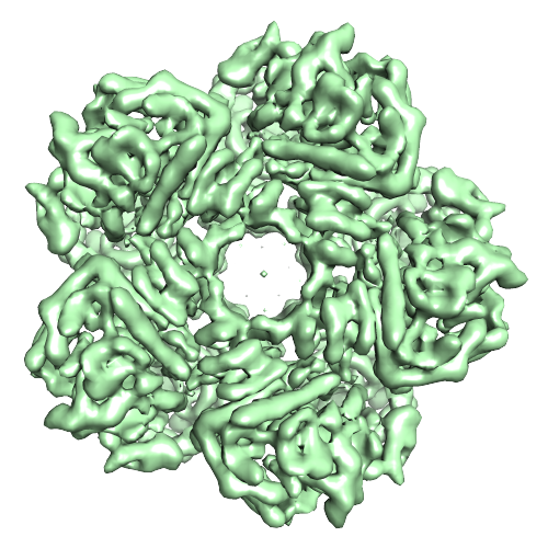 image3402.png image3402.png | 277.2 KB | ||
| Archive directory |  http://ftp.pdbj.org/pub/emdb/structures/EMD-3204ftp://ftp.pdbj.org/pub/emdb/structures/EMD-3204 http://ftp.pdbj.org/pub/emdb/structures/EMD-3204ftp://ftp.pdbj.org/pub/emdb/structures/EMD-3204 | HTTPS FTP |
-Related structure data
| Related structure data | 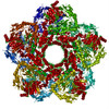 5fkxMC  3205C  3206C 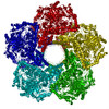 5fkzC  5fl2C M: atomic model generated by this map C: citing same article ( |
|---|---|
| Similar structure data |
-Links
| EMDB pages | EMDB (EBI/PDBe) / EMDataResource |
|---|
-Map
| File | Download / File: emd_3204.map.gz / Format: CCP4 / Size: 62.5 MB / Type: IMAGE STORED AS FLOATING POINT NUMBER (4 BYTES) | ||||||||||||||||||||||||||||||||||||||||||||||||||||||||||||
|---|---|---|---|---|---|---|---|---|---|---|---|---|---|---|---|---|---|---|---|---|---|---|---|---|---|---|---|---|---|---|---|---|---|---|---|---|---|---|---|---|---|---|---|---|---|---|---|---|---|---|---|---|---|---|---|---|---|---|---|---|---|
| Annotation | Reconstruction of active induced Lysine decarboxylase from E.coli | ||||||||||||||||||||||||||||||||||||||||||||||||||||||||||||
| Voxel size | X=Y=Z: 1.186 Å | ||||||||||||||||||||||||||||||||||||||||||||||||||||||||||||
| Density |
| ||||||||||||||||||||||||||||||||||||||||||||||||||||||||||||
| Symmetry | Space group: 1 | ||||||||||||||||||||||||||||||||||||||||||||||||||||||||||||
| Details | EMDB XML:
CCP4 map header:
| ||||||||||||||||||||||||||||||||||||||||||||||||||||||||||||
-Supplemental data
- Sample components
-Entire : E.coli Induced Lysine Decarboxylase at active pH
| Entire | Name: E.coli Induced Lysine Decarboxylase at active pH |
|---|---|
| Components |
|
-Supramolecule #1000: E.coli Induced Lysine Decarboxylase at active pH
| Supramolecule | Name: E.coli Induced Lysine Decarboxylase at active pH / type: sample / ID: 1000 / Oligomeric state: Homodecamer / Number unique components: 1 |
|---|---|
| Molecular weight | Experimental: 81.2 KDa / Theoretical: 81.2 KDa / Method: Size exclusion |
-Macromolecule #1: Inducible Lysine decarboxylase
| Macromolecule | Name: Inducible Lysine decarboxylase / type: protein_or_peptide / ID: 1 / Name.synonym: LdcI / Number of copies: 10 / Oligomeric state: Homodecamer / Recombinant expression: Yes |
|---|---|
| Source (natural) | Organism: Escherichia coli K-12 (bacteria) / Strain: K-12 / synonym: E.coli |
| Molecular weight | Experimental: 81.2 KDa / Theoretical: 81.2 KDa |
| Recombinant expression | Organism: Escherichia coli (E. coli) / Recombinant strain: MG1655 |
| Sequence | UniProtKB: Inducible lysine decarboxylase / GO: lysine catabolic process / InterPro: Ornithine/lysine/arginine decarboxylase |
-Experimental details
-Structure determination
| Method | cryo EM |
|---|---|
 Processing Processing | single particle reconstruction |
| Aggregation state | particle |
-Sample preparation
| Concentration | 2 mg/mL |
|---|---|
| Buffer | pH: 6.2 Details: 25 mM MES, 100 mM NaCl, 0.2 mM PLP, 1 mM DTT, pH 6.2 |
| Grid | Details: glow-discharged quantifoil grids 300 mesh 2/1 |
| Vitrification | Cryogen name: ETHANE / Chamber humidity: 100 % / Chamber temperature: 91 K / Instrument: FEI VITROBOT MARK III / Method: Blot for 2.5 seconds before plunging |
- Electron microscopy
| Microscope | FEI POLARA 300 |
|---|---|
| Electron beam | Acceleration voltage: 300 kV / Electron source: FIELD EMISSION GUN |
| Electron optics | Calibrated magnification: 59000 / Illumination mode: FLOOD BEAM / Imaging mode: BRIGHT FIELDBright-field microscopy / Cs: 2 mm / Nominal defocus max: 4.9 µm / Nominal defocus min: 0.6 µm / Nominal magnification: 59000 |
| Sample stage | Specimen holder: Nitrogen cooled / Specimen holder model: GATAN HELIUM |
| Temperature | Min: 90 K / Max: 92 K / Average: 91 K |
| Alignment procedure | Legacy - Electron beam tilt params: 0 |
| Date | Jun 2, 2014 |
| Image recording | Category: FILM / Film or detector model: KODAK SO-163 FILM / Digitization - Scanner: ZEISS SCAI / Digitization - Sampling interval: 7 µm / Number real images: 134 / Average electron dose: 25 e/Å2 / Bits/pixel: 8 |
| Tilt angle min | 0 |
| Tilt angle max | 0 |
| Experimental equipment |  Model: Tecnai Polara / Image courtesy: FEI Company |
-Image processing
| CTF correction | Details: each particle; full CTF correction after first peak |
|---|---|
| Final reconstruction | Applied symmetry - Point group: D5 (2x5 fold dihedral) / Algorithm: OTHER / Resolution.type: BY AUTHOR / Resolution: 6.1 Å / Resolution method: OTHER / Software - Name: RELION / Number images used: 44207 |
| Details | Reconstruction was done using RELION v1.3 with full CTF correction after first peak. |
