 Movie
Movie Controller

[English] 日本語
 Yorodumi
- EMDB-3153: Cryo-EM structure of the 60S-Arx1-Rei1 complex at near-atomic res... -
+ Open data

- Basic information
| Entry | Database: EMDB / ID: EMD-3153 | |||||||||
|---|---|---|---|---|---|---|---|---|---|---|
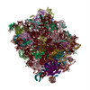
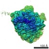
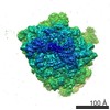
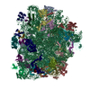
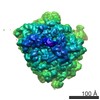
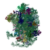
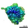
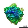
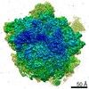
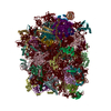
| Title | Cryo-EM structure of the 60S-Arx1-Rei1 complex at near-atomic resolution | |||||||||
 Map data Map data | Cryo-EM reconstruction of the 60S-Arx1-Rei1 complex at near-atomic resolution | |||||||||
 Sample Sample |
| |||||||||
 Keywords Keywords |  eukaryotic ribosome / 60S subunit / ribosome biogenesis / Rei1 / Arx1 / cytoplasmic maturation eukaryotic ribosome / 60S subunit / ribosome biogenesis / Rei1 / Arx1 / cytoplasmic maturation | |||||||||
| Function / homology |  Function and homology information Function and homology informationribosome biogenesis => GO:0042254 / budding cell bud growth / Hydrolases / nucleocytoplasmic transport / preribosome, large subunit precursor / ribosomal large subunit export from nucleus / ribosomal large subunit biogenesis / Neutrophil degranulation / metallopeptidase activity / mitotic cell cycle ...ribosome biogenesis => GO:0042254 / budding cell bud growth / Hydrolases / nucleocytoplasmic transport / preribosome, large subunit precursor / ribosomal large subunit export from nucleus / ribosomal large subunit biogenesis / Neutrophil degranulation / metallopeptidase activity / mitotic cell cycle / sequence-specific DNA binding / nucleolus / proteolysis / zinc ion binding / nucleoplasm / metal ion binding / nucleus / cytoplasmSimilarity search - Function | |||||||||
| Biological species |  Saccharomyces cerevisiae (brewer's yeast) Saccharomyces cerevisiae (brewer's yeast) | |||||||||
| Method | single particle reconstruction / cryo EM / Resolution: 3.7 Å | |||||||||
 Authors Authors | Greber BJ / Gerhardy S / Leitner A / Leibundgut M / Salem M / Boehringer D / Leulliot N / Aebersold R / Panse VG / Ban N | |||||||||
 Citation Citation | Journal: Cell / Year: 2016 Title: Insertion of the Biogenesis Factor Rei1 Probes the Ribosomal Tunnel during 60S Maturation. Authors: Basil Johannes Greber / Stefan Gerhardy / Alexander Leitner / Marc Leibundgut / Michèle Salem / Daniel Boehringer / Nicolas Leulliot / Ruedi Aebersold / Vikram Govind Panse / Nenad Ban /   Abstract: Eukaryotic ribosome biogenesis depends on several hundred assembly factors to produce functional 40S and 60S ribosomal subunits. The final phase of 60S subunit biogenesis is cytoplasmic maturation, ...Eukaryotic ribosome biogenesis depends on several hundred assembly factors to produce functional 40S and 60S ribosomal subunits. The final phase of 60S subunit biogenesis is cytoplasmic maturation, which includes the proofreading of functional centers of the 60S subunit and the release of several ribosome biogenesis factors. We report the cryo-electron microscopy (cryo-EM) structure of the yeast 60S subunit in complex with the biogenesis factors Rei1, Arx1, and Alb1 at 3.4 Å resolution. In addition to the network of interactions formed by Alb1, the structure reveals a mechanism for ensuring the integrity of the ribosomal polypeptide exit tunnel. Arx1 probes the entire set of inner-ring proteins surrounding the tunnel exit, and the C terminus of Rei1 is deeply inserted into the ribosomal tunnel, where it forms specific contacts along almost its entire length. We provide genetic and biochemical evidence that failure to insert the C terminus of Rei1 precludes subsequent steps of 60S maturation. | |||||||||
| History |
|
- Structure visualization
| Movie |
Movie viewer |
|---|---|
| Structure viewer | EM map: SurfViewMolmilJmol/JSmol |
| Supplemental images |

- Downloads & links
-EMDB archive
| Map data | emd_3153.map.gz | 10.1 MB | EMDB map data format | |
|---|---|---|---|---|
| Header (meta data) | emd-3153-v30.xmlemd-3153.xml | 12.8 KB 12.8 KB | Display Display | EMDB header |
| Images | 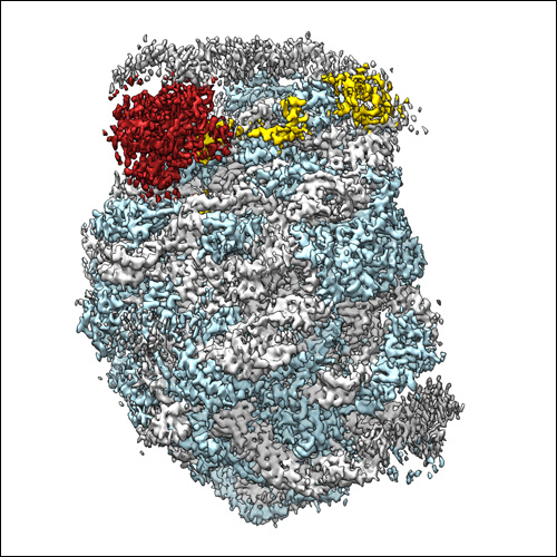 EMD_3153_500px.jpg EMD_3153_500px.jpg | 171.6 KB | ||
| Archive directory |  http://ftp.pdbj.org/pub/emdb/structures/EMD-3153ftp://ftp.pdbj.org/pub/emdb/structures/EMD-3153 http://ftp.pdbj.org/pub/emdb/structures/EMD-3153ftp://ftp.pdbj.org/pub/emdb/structures/EMD-3153 | HTTPS FTP |
-Related structure data
-Links
| EMDB pages | EMDB (EBI/PDBe) / EMDataResource |
|---|---|
| Related items in Molecule of the Month |


-Map
| File | Download / File: emd_3153.map.gz / Format: CCP4 / Size: 37.5 MB / Type: IMAGE STORED AS FLOATING POINT NUMBER (4 BYTES) | ||||||||||||||||||||||||||||||||||||||||||||||||||||||||||||||||||||
|---|---|---|---|---|---|---|---|---|---|---|---|---|---|---|---|---|---|---|---|---|---|---|---|---|---|---|---|---|---|---|---|---|---|---|---|---|---|---|---|---|---|---|---|---|---|---|---|---|---|---|---|---|---|---|---|---|---|---|---|---|---|---|---|---|---|---|---|---|---|
| Annotation | Cryo-EM reconstruction of the 60S-Arx1-Rei1 complex at near-atomic resolution | ||||||||||||||||||||||||||||||||||||||||||||||||||||||||||||||||||||
| Voxel size | X=Y=Z: 1.39 Å | ||||||||||||||||||||||||||||||||||||||||||||||||||||||||||||||||||||
| Density |
| ||||||||||||||||||||||||||||||||||||||||||||||||||||||||||||||||||||
| Symmetry | Space group: 1 | ||||||||||||||||||||||||||||||||||||||||||||||||||||||||||||||||||||
| Details | EMDB XML:
CCP4 map header:
| ||||||||||||||||||||||||||||||||||||||||||||||||||||||||||||||||||||
-Supplemental data
- Sample components
-Entire : 60S-Arx1-Rei1 complex
| Entire | Name: 60S-Arx1-Rei1 complex |
|---|---|
| Components |
|
-Supramolecule #1000: 60S-Arx1-Rei1 complex
| Supramolecule | Name: 60S-Arx1-Rei1 complex / type: sample / ID: 1000 Oligomeric state: 1:1:1 stoichiometry of 60S subunit and associated factors Number unique components: 3 |
|---|---|
| Molecular weight | Theoretical: 2.3 MDa |
-Supramolecule #1: 60S ribosomal subunit
| Supramolecule | Name: 60S ribosomal subunit / type: complex / ID: 1 / Recombinant expression: No Ribosome-details: ribosome-eukaryote: LSU 60S, LSU RNA 28S, LSU RNA 5.8S, LSU RNA 5S |
|---|---|
| Ref GO | divclassse qspanoncli ckpopupspa nclassgree n(this)spandata popltspanc lassquotlo adingbarqu otgtltimgs rcquotimgl oadinggifq uotdecodin gquotasync quotgtltsp angtdataur lajaxphp?m odetaxoamp ... divclassse qspanoncli ckpopupspa nclassgree n(this)spandata popltspanc lassquotlo adingbarqu otgtltimgs rcquotimgl oadinggifq uotdecodin gquotasync quotgtltsp angtdataur lajaxphp?m odetaxoamp kGO3A00226 25ampajax1 classpoptr giGO002262 5ispandiv |
| Source (natural) | Organism: Saccharomyces cerevisiae (brewer's yeast) / synonym: Baker's yeast / Organelle: Cytosol / Location in cell: Cytosol |
| Molecular weight | Theoretical: 2.2 MDa |
-Macromolecule #1: Arx1
| Macromolecule | Name: Arx1 / type: protein_or_peptide / ID: 1 / Number of copies: 1 / Oligomeric state: Monomer / Recombinant expression: Yes |
|---|---|
| Source (natural) | Organism: Saccharomyces cerevisiae (brewer's yeast) / Strain: BY4741 / synonym: Baker's yeast / Organelle: Cytosol and nucleus / Location in cell: Cytosol and nucleus |
| Molecular weight | Theoretical: 65 KDa |
| Recombinant expression | Organism:  Escherichia coli BL21 (bacteria) / Recombinant strain: Codon Plus / Recombinant plasmid: pET-47 Escherichia coli BL21 (bacteria) / Recombinant strain: Codon Plus / Recombinant plasmid: pET-47 |
| Sequence | UniProtKB: Probable metalloprotease ARX1 / GO: ribosome biogenesis => GO:0042254 |
-Macromolecule #2: Rei1
| Macromolecule | Name: Rei1 / type: protein_or_peptide / ID: 2 / Details: C-terminal His6-tag / Number of copies: 1 / Oligomeric state: Monomer / Recombinant expression: Yes |
|---|---|
| Source (natural) | Organism: Saccharomyces cerevisiae (brewer's yeast) / synonym: Baker's yeast / Organelle: Cytosol / Location in cell: Cytosol |
| Molecular weight | Theoretical: 45 KDa |
| Recombinant expression | Organism: Escherichia coli BL21(DE3) (bacteria) / Recombinant strain: Rosetta / Recombinant plasmid: pET-24a |
| Sequence | UniProtKB: Cytoplasmic 60S subunit biogenesis factor REI1 / GO: ribosome biogenesis => GO:0042254 |
-Experimental details
-Structure determination
| Method | cryo EM |
|---|---|
 Processing Processing | single particle reconstruction |
| Aggregation state | particle |
-Sample preparation
| Concentration | 0.2 mg/mL |
|---|---|
| Buffer | pH: 8 Details: 20 mM HEPES-KOH pH 8, 100 mM NaCl, 5 mM MgCl2, 5 mM beta-mercaptoethanol |
| Grid | Details: Quantifoil holey carbon R2/1, glow discharged |
| Vitrification | Cryogen name: ETHANE-PROPANE MIXTURE / Chamber temperature: 80 K / Instrument: HOMEMADE PLUNGER / Method: Manual blotting from the back before plunging. |
- Electron microscopy
| Microscope | FEI TITAN KRIOS |
|---|---|
| Electron beam | Acceleration voltage: 300 kV / Electron source: FIELD EMISSION GUN |
| Electron optics | Calibrated magnification: 100720 / Illumination mode: FLOOD BEAM / Imaging mode: BRIGHT FIELDBright-field microscopy / Cs: 2.7 mm / Nominal defocus max: 2.8 µm / Nominal defocus min: 0.8 µm / Nominal magnification: 59000 |
| Sample stage | Specimen holder model: FEI TITAN KRIOS AUTOGRID HOLDER |
| Details | Data collected using 4 exposures per ice hole (2 x 2 pattern). Movie mode readout in FEI EPU: 7 frames per exosure |
| Date | Jun 13, 2014 |
| Image recording | Category: CCD / Film or detector model: FEI FALCON II (4k x 4k) / Digitization - Sampling interval: 14 µm / Number real images: 1534 / Average electron dose: 20 e/Å2 Details: movie mode readout in FEI EPU: 7 frames per exposure |
| Experimental equipment |  Model: Titan Krios / Image courtesy: FEI Company |
-Image processing
| CTF correction | Details: per micrograph |
|---|---|
| Final reconstruction | Applied symmetry - Point group: C1 (asymmetric) / Algorithm: OTHER / Resolution.type: BY AUTHOR / Resolution: 3.7 Å / Resolution method: OTHER / Software - Name: CTFFIND3, RELION1.3 / Details: Particles selected in BATCHBOXER (EMAN1.9). / Number images used: 54775 |
| Details | Particles were selected semi-automatically using BATCHBOXER (EMAN). CTF correction using CTFFIND3. Reconstruction using RELION. |
-Atomic model buiding 1
| Initial model | PDB ID: |
|---|---|
| Software | Name: UCSF Chimera |
| Details | The coordinate model of the lower-resolution structure of the 60S-Arx1-Rei1 complex was fitted into the cryo-EM density using UCSF CHIMERA. The model was then adjusted using COOT (RNA) and O (proteins). |
| Refinement | Space: REAL / Protocol: RIGID BODY FIT |

