 Movie
Movie Controller

[English] 日本語
 Yorodumi
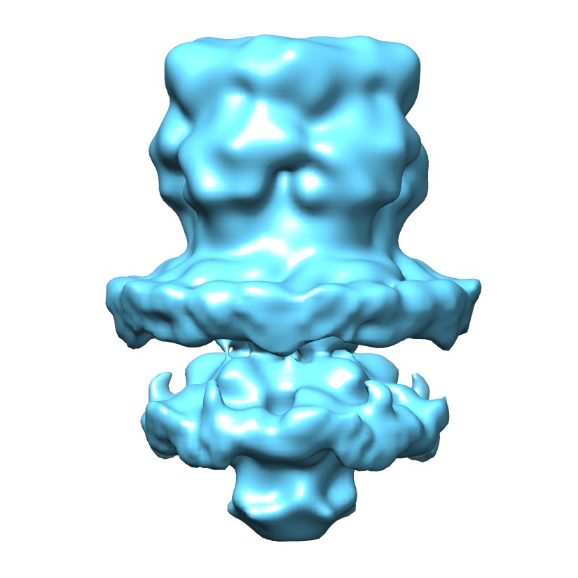
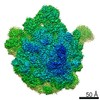
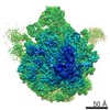
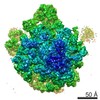
- EMDB-3108: Structure of the mouse serotonin receptor 5-HT3 in lipid vesicles -
+ Open data

- Basic information
| Entry | Database: EMDB / ID: EMD-3108 | |||||||||
|---|---|---|---|---|---|---|---|---|---|---|
| Title | Structure of the mouse serotonin receptor 5-HT3 in lipid vesicles | |||||||||
 Map data Map data | Reconstruction of the mouse 5-HT3 receptor in lipid vesicles by sub-tomogram averaging. | |||||||||
 Sample Sample |
| |||||||||
| Function / homology |  Function and homology information Function and homology informationNeurotransmitter receptors and postsynaptic signal transmission / serotonin-gated monoatomic cation channel activity / serotonin-activated cation-selective channel complex / serotonin receptor signaling pathway /  serotonin binding / acetylcholine-gated monoatomic cation-selective channel activity / inorganic cation transmembrane transport / cleavage furrow / ligand-gated monoatomic ion channel activity involved in regulation of presynaptic membrane potential / presynaptic membrane ...Neurotransmitter receptors and postsynaptic signal transmission / serotonin-gated monoatomic cation channel activity / serotonin-activated cation-selective channel complex / serotonin receptor signaling pathway / serotonin binding / acetylcholine-gated monoatomic cation-selective channel activity / inorganic cation transmembrane transport / cleavage furrow / ligand-gated monoatomic ion channel activity involved in regulation of presynaptic membrane potential / presynaptic membrane / postsynaptic membrane / neuron projection / axon / neuronal cell body / glutamatergic synapse / synapse / identical protein binding serotonin binding / acetylcholine-gated monoatomic cation-selective channel activity / inorganic cation transmembrane transport / cleavage furrow / ligand-gated monoatomic ion channel activity involved in regulation of presynaptic membrane potential / presynaptic membrane ...Neurotransmitter receptors and postsynaptic signal transmission / serotonin-gated monoatomic cation channel activity / serotonin-activated cation-selective channel complex / serotonin receptor signaling pathway / serotonin binding / acetylcholine-gated monoatomic cation-selective channel activity / inorganic cation transmembrane transport / cleavage furrow / ligand-gated monoatomic ion channel activity involved in regulation of presynaptic membrane potential / presynaptic membrane / postsynaptic membrane / neuron projection / axon / neuronal cell body / glutamatergic synapse / synapse / identical protein bindingSimilarity search - Function | |||||||||
| Biological species |  Mus musculus (house mouse) Mus musculus (house mouse) | |||||||||
| Method | subtomogram averaging / cryo EM / negative staining / Resolution: 12.0 Å | |||||||||
 Authors Authors | Kudryashev M / Castano-Diez D / Deluz C / Hassaine G / Graf-Meyer A / Vogel H / Stahlberg H | |||||||||
 Citation Citation | Journal: Structure / Year: 2016 Title: The Structure of the Mouse Serotonin 5-HT3 Receptor in Lipid Vesicles. Authors: Mikhail Kudryashev / Daniel Castaño-Díez / Cédric Deluz / Gherici Hassaine / Luigino Grasso / Alexandra Graf-Meyer / Horst Vogel / Henning Stahlberg /   Abstract: The function of membrane proteins is best understood if their structure in the lipid membrane is known. Here, we determined the structure of the mouse serotonin 5-HT3 receptor inserted in lipid ...The function of membrane proteins is best understood if their structure in the lipid membrane is known. Here, we determined the structure of the mouse serotonin 5-HT3 receptor inserted in lipid bilayers to a resolution of 12 Å without stabilizing antibodies by cryo electron tomography and subtomogram averaging. The reconstruction reveals protein secondary structure elements in the transmembrane region, the extracellular pore, and the transmembrane channel pathway, showing an overall similarity to the available X-ray model of the truncated 5-HT3 receptor determined in the presence of a stabilizing nanobody. Structural analysis of the 5-HT3 receptor embedded in a lipid bilayer allowed the position of the membrane to be determined. Interactions between the densely packed receptors in lipids were visualized, revealing that the interactions were maintained by the short horizontal helices. In combination with methodological improvements, our approach enables the structural analysis of membrane proteins in response to voltage and ligand gating. | |||||||||
| History |
|
- Structure visualization
| Movie |
Movie viewer |
|---|---|
| Structure viewer | EM map: SurfViewMolmilJmol/JSmol |
| Supplemental images |
- Downloads & links
-EMDB archive
| Map data | emd_3108.map.gz | 7.2 MB | EMDB map data format | |
|---|---|---|---|---|
| Header (meta data) | emd-3108-v30.xmlemd-3108.xml | 11.7 KB 11.7 KB | Display Display | EMDB header |
| Images | EMD-3108.tif | 327.3 KB | ||
| Masks | emd_3108_msk_1.map | 8 MB | Mask map | |
| Archive directory |  http://ftp.pdbj.org/pub/emdb/structures/EMD-3108ftp://ftp.pdbj.org/pub/emdb/structures/EMD-3108 http://ftp.pdbj.org/pub/emdb/structures/EMD-3108ftp://ftp.pdbj.org/pub/emdb/structures/EMD-3108 | HTTPS FTP |
-Related structure data
| Similar structure data | |
|---|---|
| EM raw data | EMPIAR-10046 (Title: Cryo electron tomography of mouse 5-HT3 receptors in lipid vesicles Data size: 158.8 Data #1: Tilt series of the 5-HT3 receptors reconstituted to lipid vesicles [class averages]) |
-Links
| EMDB pages | EMDB (EBI/PDBe) / EMDataResource |
|---|---|
| Related items in Molecule of the Month |


-Map
| File | Download / File: emd_3108.map.gz / Format: CCP4 / Size: 7.8 MB / Type: IMAGE STORED AS FLOATING POINT NUMBER (4 BYTES) | ||||||||||||||||||||||||||||||||||||||||||||||||||||||||||||||||||||
|---|---|---|---|---|---|---|---|---|---|---|---|---|---|---|---|---|---|---|---|---|---|---|---|---|---|---|---|---|---|---|---|---|---|---|---|---|---|---|---|---|---|---|---|---|---|---|---|---|---|---|---|---|---|---|---|---|---|---|---|---|---|---|---|---|---|---|---|---|---|
| Annotation | Reconstruction of the mouse 5-HT3 receptor in lipid vesicles by sub-tomogram averaging. | ||||||||||||||||||||||||||||||||||||||||||||||||||||||||||||||||||||
| Voxel size | X=Y=Z: 1.67 Å | ||||||||||||||||||||||||||||||||||||||||||||||||||||||||||||||||||||
| Density |
| ||||||||||||||||||||||||||||||||||||||||||||||||||||||||||||||||||||
| Symmetry | Space group: 1 | ||||||||||||||||||||||||||||||||||||||||||||||||||||||||||||||||||||
| Details | EMDB XML:
CCP4 map header:
| ||||||||||||||||||||||||||||||||||||||||||||||||||||||||||||||||||||
-Supplemental data
-Segmentation: mask for the final visualization, contains one pentamer
| Annotation | mask for the final visualization, contains one pentamer | ||||||||||||
|---|---|---|---|---|---|---|---|---|---|---|---|---|---|
| File | emd_3108_msk_1.map | ||||||||||||
| Projections & Slices |
| ||||||||||||
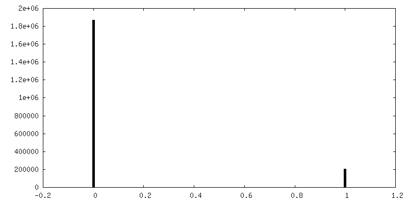
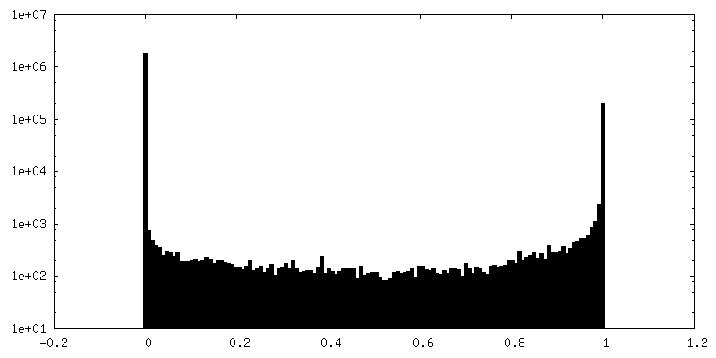
| Density Histograms |
 Z
Z Y
Y X
X


- Sample components
-Entire : Mouse serotonin receptor 5-HT3
| Entire | Name: Mouse serotonin receptor 5-HT3 |
|---|---|
| Components |
|
-Supramolecule #1000: Mouse serotonin receptor 5-HT3
| Supramolecule | Name: Mouse serotonin receptor 5-HT3 / type: sample / ID: 1000 / Oligomeric state: pentamer / Number unique components: 1 |
|---|---|
| Molecular weight | Theoretical: 244 MDa |
-Macromolecule #1: 5-hydroxytryptamine receptor
| Macromolecule | Name: 5-hydroxytryptamine receptor / type: protein_or_peptide / ID: 1 / Name.synonym: 5-HT3 / Details: Protein reconstituted into lipid vesicles. / Number of copies: 5 / Oligomeric state: pentamer / Recombinant expression: Yes |
|---|---|
| Source (natural) | Organism: Mus musculus (house mouse) / synonym: Mouse / Location in cell: Plasma membrane |
| Molecular weight | Theoretical: 244 MDa |
| Recombinant expression | Organism:  Homo sapiens (human) / Recombinant cell: T-REx-293 cells / Recombinant plasmid: pcDNA5/TO Homo sapiens (human) / Recombinant cell: T-REx-293 cells / Recombinant plasmid: pcDNA5/TO |
| Sequence | UniProtKB: 5-hydroxytryptamine receptor 3A |
-Experimental details
-Structure determination
| Method | negative staining, cryo EM |
|---|---|
 Processing Processing | subtomogram averaging |
| Aggregation state | particle |
-Sample preparation
| Staining | Type: NEGATIVE / Details: Cryo-preparation |
|---|---|
| Grid | Details: Holey carbon grids |
| Vitrification | Cryogen name: ETHANE / Chamber humidity: 90 % / Chamber temperature: 90 K / Instrument: LEICA EM GP / Method: Blot for 2 second before plunding |
- Electron microscopy
| Microscope | FEI TITAN KRIOS |
|---|---|
| Electron beam | Acceleration voltage: 300 kV / Electron source: FIELD EMISSION GUN |
| Electron optics | Illumination mode: FLOOD BEAM / Imaging mode: BRIGHT FIELDBright-field microscopy / Cs: 2.7 mm / Nominal defocus max: 4.5 µm / Nominal defocus min: 2.5 µm |
| Sample stage | Specimen holder model: FEI TITAN KRIOS AUTOGRID HOLDER / Tilt series - Axis1 - Min angle: -60 ° / Tilt series - Axis1 - Max angle: 60 ° |
| Alignment procedure | Legacy - Astigmatism: corrected at high magnification |
| Details | tomograms acquired without the energy filtration |
| Date | Dec 1, 2013 |
| Image recording | Category: CCD / Film or detector model: GATAN K2 (4k x 4k) / Number real images: 46 / Average electron dose: 41 e/Å2 / Details: 46 tomograms were selected for processing |
| Experimental equipment |  Model: Titan Krios / Image courtesy: FEI Company |
-Image processing
| CTF correction | Details: ctfplotter |
|---|---|
| Final 3D classification | Number classes: 20 |
| Final reconstruction | Applied symmetry - Point group: C5 (5 fold cyclic) / Resolution.type: BY AUTHOR / Resolution: 12.0 Å / Resolution method: OTHER / Software - Name: IMOD, Dynamo / Number subtomograms used: 16000 |
| Details | Particles were automatically picked from the surface of the vesicles in CTF-corrected cryo-electron tomograms. After a rough alignment deep classification into 20 classes was performed including 4 neighbouring pentamers in the alignment mask. Finally the central pentamers from each class were cropped, aligned and averaged resulting in a final structure. |
