- EMDB-3034: Structure of the 26S proteasome-Ubp6 complex -
+
Open data
ID or keywords:
Loading...
-
Basic information
Entry
Database: EMDB / ID: EMD-3034
Title
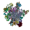
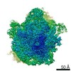
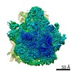
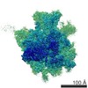
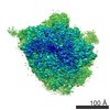
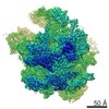
Structure of the 26S proteasome-Ubp6 complex
Map data
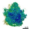
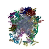
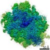
Reconstruction of the 26S proteasome in presence of Ubp6 and ubiquitin aldehyde
Sample
Sample: 26S Proteasome from Saccharomyces cerevisiae in the presence of Saccharomyces cerevisiae Ubp6 and ubiquitin aldehydeProteasome
Protein or peptide: 26S ProteasomeProteasome
Protein or peptide: Ubp6
Protein or peptide: ubiqutin aldehyde
Keywords
conformational switching / protein degradation / proteostasis / quality control / Ubp6
Function / homology
Function and homology information
SAGA complex localization to transcription regulatory region / mitochondria-associated ubiquitin-dependent protein catabolic process / peroxisome fission / negative regulation of proteasomal protein catabolic process / regulation of proteasomal ubiquitin-dependent protein catabolic process / proteasome storage granule assembly / transcription export complex 2 / proteasome regulatory particle assembly / protein deneddylation / nonfunctional rRNA decay ...SAGA complex localization to transcription regulatory region / mitochondria-associated ubiquitin-dependent protein catabolic process / peroxisome fission / negative regulation of proteasomal protein catabolic process / regulation of proteasomal ubiquitin-dependent protein catabolic process / proteasome storage granule assembly / transcription export complex 2 / proteasome regulatory particle assembly / protein deneddylation / nonfunctional rRNA decay / maintenance of DNA trinucleotide repeats / filamentous growth / COP9 signalosome / proteasome regulatory particle / cytosolic proteasome complex / proteasome regulatory particle, lid subcomplex / proteasome-activating activity / protein-containing complex localization / mitochondrial fission / proteasome regulatory particle, base subcomplex / K48-linked polyubiquitin modification-dependent protein binding / proteasome core complex assembly / nuclear outer membrane-endoplasmic reticulum membrane network / Cross-presentation of soluble exogenous antigens (endosomes) / TNFR2 non-canonical NF-kB pathway / proteasomal ubiquitin-independent protein catabolic process / Ub-specific processing proteases / peptide catabolic process / proteasome binding / regulation of protein catabolic process / protein deubiquitination / Peptide chain elongation / Selenocysteine synthesis / proteasome storage granule / Formation of a pool of free 40S subunits / Eukaryotic Translation Termination / Response of EIF2AK4 (GCN2) to amino acid deficiency / SRP-dependent cotranslational protein targeting to membrane / Viral mRNA Translation / polyubiquitin modification-dependent protein binding / Nonsense Mediated Decay (NMD) independent of the Exon Junction Complex (EJC) / endopeptidase activator activity / GTP hydrolysis and joining of the 60S ribosomal subunit / L13a-mediated translational silencing of Ceruloplasmin expression / Major pathway of rRNA processing in the nucleolus and cytosol / proteasome assembly / positive regulation of RNA polymerase II transcription preinitiation complex assembly / proteasome endopeptidase complex / proteasome core complex, beta-subunit complex / proteasome core complex, alpha-subunit complex / threonine-type endopeptidase activity / Nonsense Mediated Decay (NMD) enhanced by the Exon Junction Complex (EJC) / regulation of proteasomal protein catabolic process / enzyme regulator activity / mRNA export from nucleus / : / protein folding chaperone / Maturation of protein E / Maturation of protein E / ER Quality Control Compartment (ERQC) / Myoclonic epilepsy of Lafora / FLT3 signaling by CBL mutants / Prevention of phagosomal-lysosomal fusion / IRAK2 mediated activation of TAK1 complex / Alpha-protein kinase 1 signaling pathway / Glycogen synthesis / IRAK1 recruits IKK complex / IRAK1 recruits IKK complex upon TLR7/8 or 9 stimulation / Membrane binding and targetting of GAG proteins / Constitutive Signaling by NOTCH1 HD Domain Mutants / Endosomal Sorting Complex Required For Transport (ESCRT) / NOTCH2 Activation and Transmission of Signal to the Nucleus / IRAK2 mediated activation of TAK1 complex upon TLR7/8 or 9 stimulation / PTK6 Regulates RTKs and Their Effectors AKT1 and DOK1 / Negative regulation of FLT3 / Regulation of FZD by ubiquitination / TICAM1,TRAF6-dependent induction of TAK1 complex / TICAM1-dependent activation of IRF3/IRF7 / APC/C:Cdc20 mediated degradation of Cyclin B / Neutrophil degranulation / Downregulation of ERBB4 signaling / p75NTR recruits signalling complexes / TRAF6 mediated IRF7 activation in TLR7/8 or 9 signaling / APC-Cdc20 mediated degradation of Nek2A / PINK1-PRKN Mediated Mitophagy / TRAF6-mediated induction of TAK1 complex within TLR4 complex / InlA-mediated entry of Listeria monocytogenes into host cells / Pexophagy / Regulation of innate immune responses to cytosolic DNA / VLDLR internalisation and degradation / Downregulation of ERBB2:ERBB3 signaling / NRIF signals cell death from the nucleus / Activated NOTCH1 Transmits Signal to the Nucleus / Translesion synthesis by REV1 / NF-kB is activated and signals survival / Regulation of PTEN localization / Translesion synthesis by POLK / Regulation of BACH1 activity / Synthesis of active ubiquitin: roles of E1 and E2 enzymes / proteasome complex Similarity search - Function
Journal: Proc Natl Acad Sci U S A / Year: 2015 Title: Structural characterization of the interaction of Ubp6 with the 26S proteasome. Authors: Antje Aufderheide / Florian Beck / Florian Stengel / Michaela Hartwig / Andreas Schweitzer / Günter Pfeifer / Alfred L Goldberg / Eri Sakata / Wolfgang Baumeister / Friedrich Förster / Abstract: In eukaryotic cells, the 26S proteasome is responsible for the regulated degradation of intracellular proteins. Several cofactors interact transiently with this large macromolecular machine and ...In eukaryotic cells, the 26S proteasome is responsible for the regulated degradation of intracellular proteins. Several cofactors interact transiently with this large macromolecular machine and modulate its function. The deubiquitylating enzyme ubiquitin C-terminal hydrolase 6 [Ubp6; ubiquitin-specific protease (USP) 14 in mammals] is the most abundant proteasome-interacting protein and has multiple roles in regulating proteasome function. Here, we investigate the structural basis of the interaction between Ubp6 and the 26S proteasome in the presence and absence of the inhibitor ubiquitin aldehyde. To this end we have used single-particle electron cryomicroscopy in combination with cross-linking and mass spectrometry. Ubp6 binds to the regulatory particle non-ATPase (Rpn) 1 via its N-terminal ubiquitin-like domain, whereas its catalytic USP domain is positioned variably. Addition of ubiquitin aldehyde stabilizes the binding of the USP domain in a position where it bridges the proteasome subunits Rpn1 and the regulatory particle triple-A ATPase (Rpt) 1. The USP domain binds to Rpt1 in the immediate vicinity of the Ubp6 active site, which may effect its activation. The catalytic triad is positioned in proximity to the mouth of the ATPase module and to the deubiquitylating enzyme Rpn11, strongly implying their functional linkage. On the proteasome side, binding of Ubp6 favors conformational switching of the 26S proteasome into an intermediate-energy conformational state, in particular upon the addition of ubiquitin aldehyde. This modulation of the conformational space of the 26S proteasome by Ubp6 explains the effects of Ubp6 on the kinetics of proteasomal degradation.
History
Deposition
Jun 1, 2015
-
Header (metadata) release
Jul 8, 2015
-
Map release
Jul 15, 2015
-
Update
Aug 12, 2015
-
Current status
Aug 12, 2015
Processing site: PDBe / Status: Released
-
Structure visualization
Movie
Surface view with section colored by density value
#112 - Apr 2009 Oct and Sox Transcription Factors similarity (1)
-
Map
File
Download / File: emd_3034.map.gz / Format: CCP4 / Size: 81.8 MB / Type: IMAGE STORED AS FLOATING POINT NUMBER (4 BYTES)
Annotation
Reconstruction of the 26S proteasome in presence of Ubp6 and ubiquitin aldehyde
Voxel size
X=Y=Z: 1.99 Å
Density
Contour Level
By AUTHOR: 1.17 / Movie #1: 1.17
Minimum - Maximum
-11.709941860000001 - 12.581256870000001
Average (Standard dev.)
0.00614737 (±0.297102)
Symmetry
Space group: 1
Details
EMDB XML:
Map geometry
Axis order
X
Y
Z
Origin
0
0
0
Dimensions
280
280
280
Spacing
280
280
280
Cell
A=B=C: 557.2 Å α=β=γ: 90.0 °
CCP4 map header:
mode
Image stored as Reals
Å/pix. X/Y/Z
1.99
1.99
1.99
M x/y/z
280
280
280
origin x/y/z
0.000
0.000
0.000
length x/y/z
557.200
557.200
557.200
α/β/γ
90.000
90.000
90.000
start NX/NY/NZ
-147
-147
-146
NX/NY/NZ
294
294
294
MAP C/R/S
1
2
3
start NC/NR/NS
0
0
0
NC/NR/NS
280
280
280
D min/max/mean
-11.710
12.581
0.006
-
Supplemental data
-
Sample components
-
Entire : 26S Proteasome from Saccharomyces cerevisiae in the presence of S...
Entire
Name: 26S Proteasome from Saccharomyces cerevisiae in the presence of Saccharomyces cerevisiae Ubp6 and ubiquitin aldehydeProteasome
Components
Sample: 26S Proteasome from Saccharomyces cerevisiae in the presence of Saccharomyces cerevisiae Ubp6 and ubiquitin aldehydeProteasome
Protein or peptide: 26S ProteasomeProteasome
Protein or peptide: Ubp6
Protein or peptide: ubiqutin aldehyde
-
Supramolecule #1000: 26S Proteasome from Saccharomyces cerevisiae in the presence of S...
Supramolecule
Name: 26S Proteasome from Saccharomyces cerevisiae in the presence of Saccharomyces cerevisiae Ubp6 and ubiquitin aldehyde type: sample / ID: 1000 / Number unique components: 3
Category: CCD / Film or detector model: FEI FALCON II (4k x 4k) / Number real images: 5630 / Average electron dose: 45 e/Å2 / Details: Every image is the average of 7 aligned frames.
Tilt angle min
0
Tilt angle max
0
Experimental equipment
Model: Titan Krios / Image courtesy: FEI Company
-
Image processing
CTF correction
Details: micrograph
Final two d classification
Number classes: 29100
Final reconstruction
Applied symmetry - Point group: C1 (asymmetric) / Algorithm: OTHER / Resolution.type: BY AUTHOR / Resolution: 9.5 Å / Resolution method: OTHER / Software - Name: xmipp / Number images used: 53000
Details
The particles were selected using an automatic selection program. Each physical 26S particles was considered as two particles for processing according to pseudo-C2 symmetry.
+
About Yorodumi
-
News
-
Feb 9, 2022. New format data for meta-information of EMDB entries
New format data for meta-information of EMDB entries
Version 3 of the EMDB header file is now the official format.
The previous official version 1.9 will be removed from the archive.
In the structure databanks used in Yorodumi, some data are registered as the other names, "COVID-19 virus" and "2019-nCoV". Here are the details of the virus and the list of structure data.
Jan 31, 2019. EMDB accession codes are about to change! (news from PDBe EMDB page)
EMDB accession codes are about to change! (news from PDBe EMDB page)
The allocation of 4 digits for EMDB accession codes will soon come to an end. Whilst these codes will remain in use, new EMDB accession codes will include an additional digit and will expand incrementally as the available range of codes is exhausted. The current 4-digit format prefixed with “EMD-” (i.e. EMD-XXXX) will advance to a 5-digit format (i.e. EMD-XXXXX), and so on. It is currently estimated that the 4-digit codes will be depleted around Spring 2019, at which point the 5-digit format will come into force.
The EM Navigator/Yorodumi systems omit the EMD- prefix.
Related info.:Q: What is EMD? / ID/Accession-code notation in Yorodumi/EM Navigator
Yorodumi is a browser for structure data from EMDB, PDB, SASBDB, etc.
This page is also the successor to EM Navigator detail page, and also detail information page/front-end page for Omokage search.
The word "yorodu" (or yorozu) is an old Japanese word meaning "ten thousand". "mi" (miru) is to see.
Related info.:EMDB / PDB / SASBDB / Comparison of 3 databanks / Yorodumi Search / Aug 31, 2016. New EM Navigator & Yorodumi / Yorodumi Papers / Jmol/JSmol / Function and homology information / Changes in new EM Navigator and Yorodumi
 Movie
Movie Controller


 Open data
 Basic information
 Map data
 Sample
 Keywords
 protein degradation /
  Function and homology information

 Authors
 Citation

 Structure visualization
 Downloads & links
 http://ftp.pdbj.org/pub/emdb/structures/EMD-3034
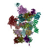


 Sample components
 Processing
 Electron microscopy
